

Antiviral Effects of Tecovirimat and Cellular Ultrastructural Changes in Human Bronchial Epithelial Cell Line Following Monkeypox Virus Infection
- PMID: 40141361
- PMCID: PMC11942983
- DOI: 10.3390/ijms26062718
Antiviral Effects of Tecovirimat and Cellular Ultrastructural Changes in Human Bronchial Epithelial Cell Line Following Monkeypox Virus Infection
Abstract
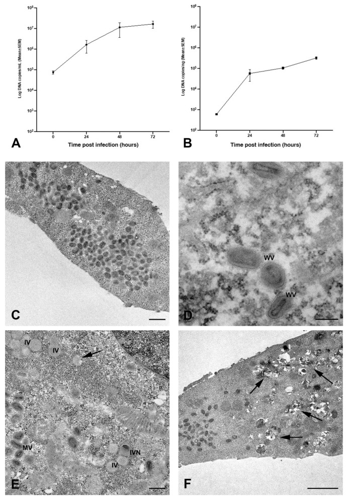
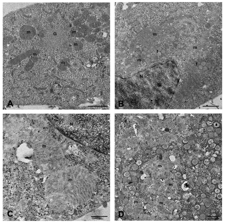
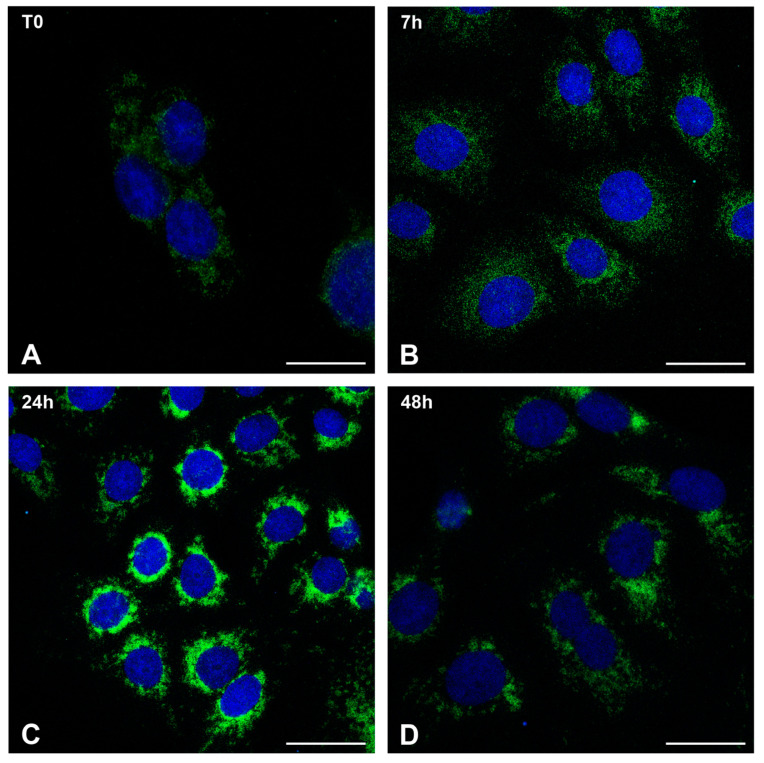
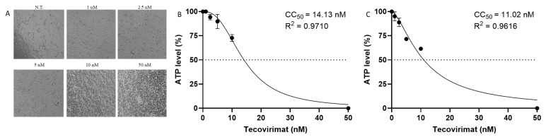
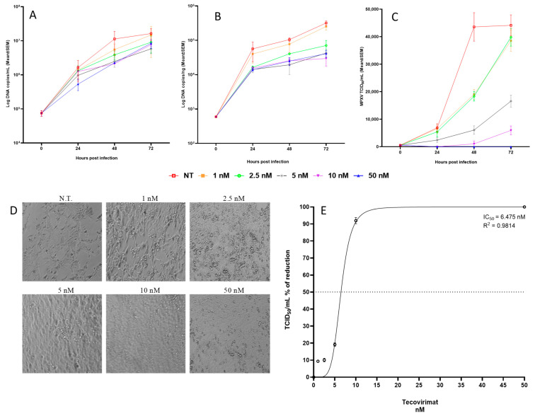
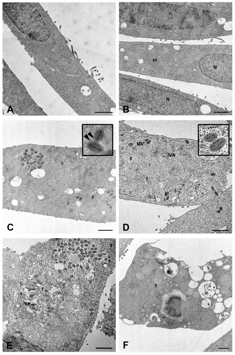
The mpox virus (MPXV) Clade IIb outbreak in 2022 was the biggest one ever to occur outside Africa, causing different types of clinical symptoms and levels of disease severity. There is no currently approved treatment for mpox, but Tecovirimat has proven effective against known orthopoxviruses in several animal models and Vero cell cultures. Since serious complications, including lung involvement, have been reported, especially in immunocompromised people, we investigated the effects of MPXV infection on the in vitro model of lung airway epithelium (Calu-3 cell line) and examined MPXV replication kinetic and related ultrastructural changes, also performing dose-response studies to measure Tecovirimat antiviral activity. Our results highlighted an active replication of MPXV in Calu-3 cells linked to mitochondrial structural modifications with perinuclear relocation and the formation of cytoplasmic vacuoles. Treatment with Tecovirimat consistently reduced viral replication both in supernatants (81%) and inside cells (77%) and ultimately stopped viral infectivity (92% of cytopathic effect reduction) after 48 h of infection. Drug administration inhibited the final wrapping of mature viral particles, causing extensive cytoplasmic vacuolation. Our results demonstrated Tecovirimat's in vitro effectiveness against MPXV at the nanomolar concentration on Calu-3 cells. This suggests a potential rationale for using this drug for patients with mpox severe disease and lung involvement.
Keywords: Calu-3 cells; MPXV; antiviral activity; electron microscopy; mitochondria; mpox disease; tecovirimat.
Conflict of interest statement
The authors declare that they have no known competing financial interests or personal relationships that could have appeared to influence the work reported in this paper.
Figures
Similar articles
-
Emerging variants of Mpox virus and tecovirimat resistance: Genomic insights and implications for treatment strategies.Virology. 2025 Jul;608:110532. doi: 10.1016/j.virol.2025.110532. Epub 2025 Apr 12. Virology. 2025. PMID: 40245474 Review.
-
Functional characteristics of plitidepsin as an antiviral treatment against monkeypox virus infection.Antiviral Res. 2025 Sep;241:106238. doi: 10.1016/j.antiviral.2025.106238. Epub 2025 Jul 15. Antiviral Res. 2025. PMID: 40675496
-
Treatment efficacy of cidofovir and brincidofovir against clade II Monkeypox virus isolates.Antiviral Res. 2024 Nov;231:105995. doi: 10.1016/j.antiviral.2024.105995. Epub 2024 Sep 5. Antiviral Res. 2024. PMID: 39243894
-
Notes from the Field: Mpox Cluster Caused by Tecovirimat-Resistant Monkeypox Virus - Five States, October 2023-February 2024.MMWR Morb Mortal Wkly Rep. 2024 Oct 10;73(40):903-905. doi: 10.15585/mmwr.mm7340a3. MMWR Morb Mortal Wkly Rep. 2024. PMID: 39388389 Free PMC article.
-
New Perspectives on Antimicrobial Agents: Tecovirimat for Treatment of Human Monkeypox Virus.Antimicrob Agents Chemother. 2022 Dec 20;66(12):e0122622. doi: 10.1128/aac.01226-22. Epub 2022 Nov 14. Antimicrob Agents Chemother. 2022. PMID: 36374026 Free PMC article. Review.
References
-
- ICoToV Virus Taxonomy: 2023 Release. [(accessed on 11 July 2023)]. Available online: https://talk.ictvonline.org/taxonomy.
-
- Adler H., Gould S., Hine P., Snell L.B., Wong W., Houlihan C.F., Osborne J.C., Rampling T., Beadsworth M.B., Duncan C.J., et al. Clinical features and management of human monkeypox: A retrospective observational study in the UK. Lancet Infect. Dis. 2022;22:1153–1162. doi: 10.1016/S1473-3099(22)00228-6. - DOI - PMC - PubMed
-
- World Health Organization Mpox (Monkeypox) Outbreak 2022-Global. 2022. [(accessed on 11 July 2023)]. Available online: https://www.who.int/emergencies/situations/monkeypox-oubreak-2022.
-
- Antinori A., Mazzotta V., Vita S., Carletti F., Tacconi D., Lapini L.E., D’Abramo A., Cicalini S., Lapa D., Pittalis S., et al. Epidemiological, clinical and virological characteristics of four cases of monkeypox support transmission through sexual contact, Italy, May 2022. Euro. Surveill. 2022;27:2200421. doi: 10.2807/1560-7917.ES.2022.27.22.2200421. - DOI - PMC - PubMed
MeSH terms
Substances
Grants and funding
LinkOut - more resources
Full Text Sources
