. 2025 Mar 19;26(6):2769.
doi: 10.3390/ijms26062769.

Pulmonary Myeloid Cells in Mild Cases of COVID-19 Upregulate the Intracellular Fc Receptor TRIM21 and Transcribe Proteasome-Associated Molecules

Affiliations

Pulmonary Myeloid Cells in Mild Cases of COVID-19 Upregulate the Intracellular Fc Receptor TRIM21 and Transcribe Proteasome-Associated Molecules

Andrea Henriques-Pons et al. Int J Mol Sci. .

Abstract

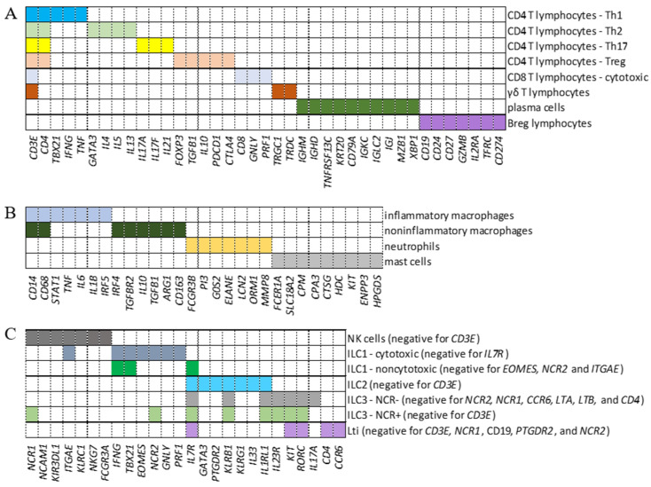
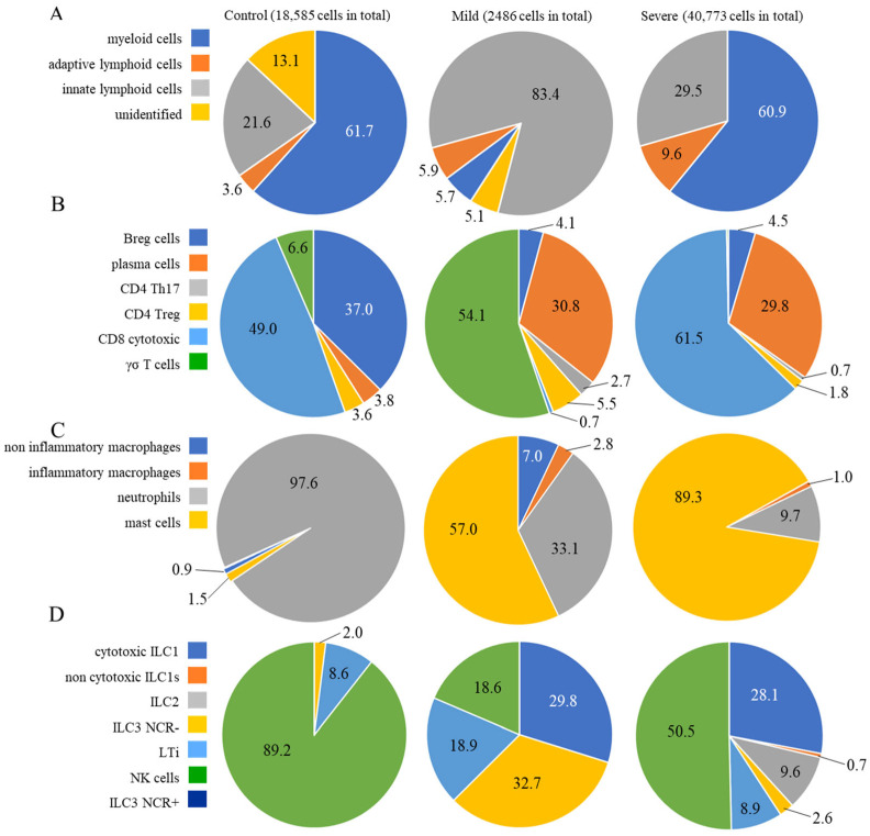
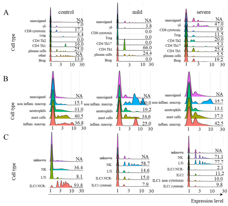
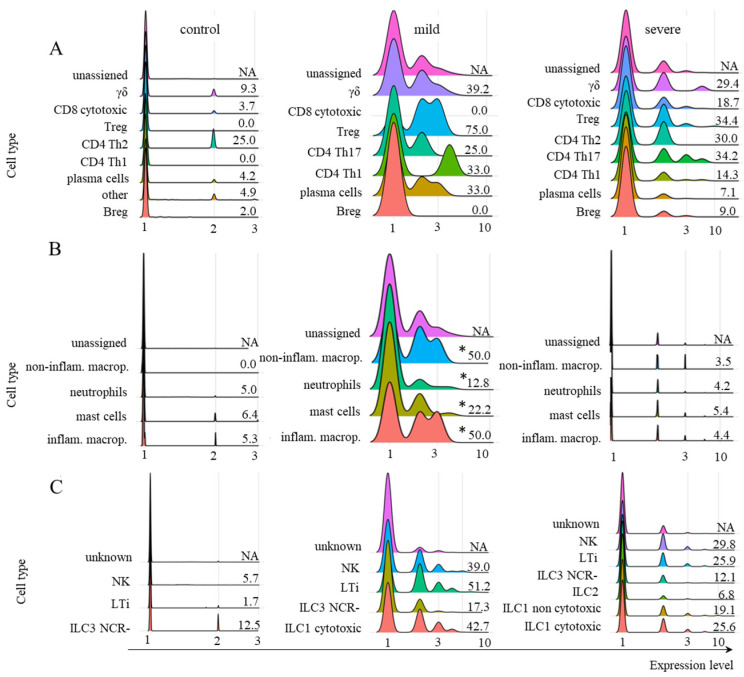
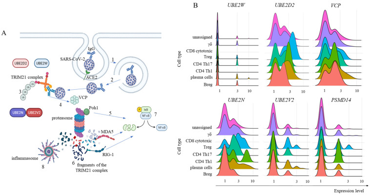
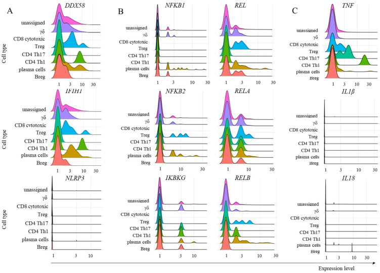
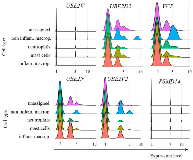
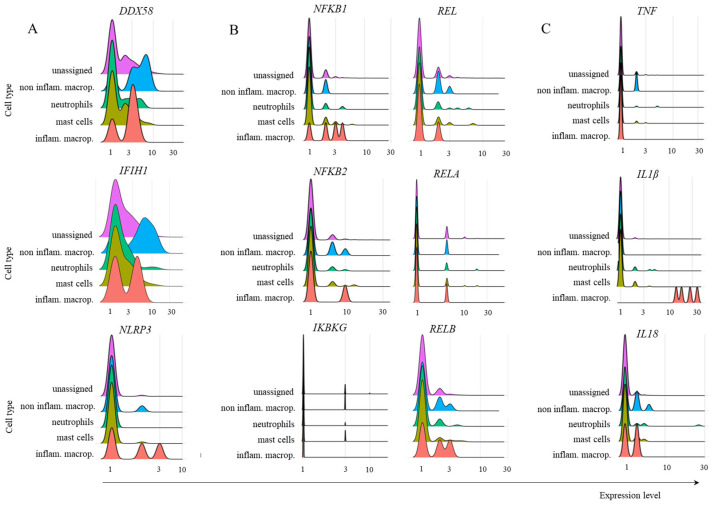
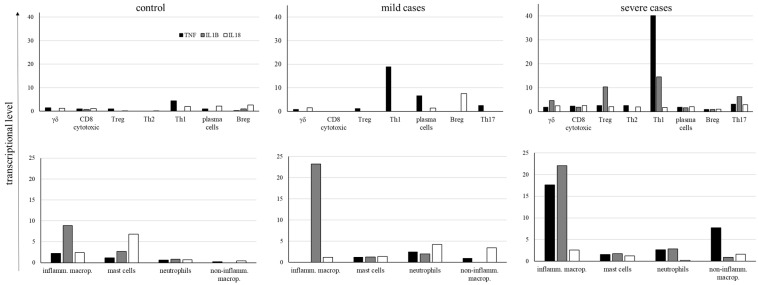
Much remains to be understood about COVID-19, but the protective role of antibodies (Igs) is widely accepted in SARS-CoV-2 infection. Igs' functions are mainly carried out by receptors that bind to their Fc portion (FcR), and less attention has been dedicated to the cytoplasmic members of this family. In this work, we used single-cell RNA sequencing (scRNA-seq) data to discern cell populations in bronchoalveolar lavage fluid obtained from healthy individuals and patients with mild or severe COVID-19. Then, we evaluated the transcription of neonatal FcR (FcRn, FCGRT gene) and tripartite motif-containing protein 21 (TRIM21) and its downstream signaling components. The TRIM21 pathway is vital for virus infections as it has a dual function, leading opsonized viruses to degradation by proteasomes and the activation of innate inflammatory anti-virus response. The transcriptional level of FCGRT showed no statistical differences in any cell population comparing the three groups of patients. On the other hand, TRIM21 transcription was significantly higher in myeloid cells collected from patients with mild COVID-19. When comparing mild with severe cases, there was no statistical difference in TRIM21 transcription in lung adaptive lymphoid cells and innate lymphoid cells (ILC). Yet, we analyzed the transcription of all downstream signaling molecules in myeloid and, as most cells expressed the receptor, in adaptive lymphoid cells. Moreover, ILCs from mild cases and all cell populations from severe cases were missing most downstream components of the pathway. We observed that members of the ubiquitin-proteasome system (UPS) and other components associated with TRIM21 proteasomal degradation were transcribed in mild cases. Despite the transcription of the danger sensors DDX58 and IFIH1, the transcriptional level of inflammatory IL1B and IL18 was generally very low, along with the NLRP3 danger sensor, members of the NF-κB pathway, and TNF. Therefore, our data suggest that TRIM21 may contribute to SARS-CoV-2 protection by reducing the viral load, while the inflammatory branch of the pathway would be silenced, leading to no pathogenic cytokine production.

Keywords: COVID-19; Fc receptors; TRIM21; inflammatory response; scRNA-seq.


Conflict of interest statement

The authors declare that the research was conducted without any commercial or financial relationships that could be construed as potential conflicts of interest.

Figures

Figure 1
Figure 1
Identification of cell populations. Panels (AC) show the combinations of genes used to identify adaptive lymphoid cells (A), myeloid cells (B), and innate lymphoid cells (ILCs) (C), respectively. Each colored segment shows the combination of genes that must be simultaneously transcribed to identify all indicated subpopulations. In the case of ILCs, the genes indicated in parentheses as negative represent a group of additional genes that must not be transcribed for this cellular classification.
Figure 2
Figure 2
Percentages of different cellular populations among patient groups. The percentage of each cell cluster is shown in (A), and the percentages of each subpopulation per cluster are shown in the other panels. Adaptive lymphoid cells are shown in (B); myeloid cells are shown in (C); and ILCs are shown in (D). The total number of cells analyzed per group of patients is shown at the top of the figure.
Figure 3
Figure 3
Transcriptional profile of FCGRT in different cellular populations. The transcriptional profile of FCGRT in the indicated subpopulations of adaptive lymphoid cells (A), myeloid cells (B), and ILCs (C) is shown for control individuals or patients with mild or severe symptoms of COVID-19. The information next to each population represents the percentage of cells whose FCGRT transcriptional level was above zero. The colors assigned to each histogram may vary between cell populations, and correct identification should be based on the indicated population name. No CD4+ Th2 lymphocytes were identified in the samples from mild cases. To calculate the percentage of FCGRT+ cells, we used the number of total cells per subpopulation per patient group (considered 100%). This number was revealed by the phenotypic analysis indicated in the MM section. Then, we determined the number of FCGRT+ cells per subpopulation. NA means not available. The statistical analysis returned no significant differences between any cell subpopulation and patient group (considering p ≤ 0.05).
Figure 4
Figure 4
Transcriptional profile of TRIM21 in different cell populations. The transcriptional profile of TRIM21 is shown for adaptive lymphoid cells (A), myeloid cells (B), and ILCs (C). The samples were obtained from healthy control individuals or patients with mild or severe symptoms of COVID-19. The information next to each subpopulation represents the percentage of cells with TRIM21 transcriptional levels above zero. To calculate the percentages, we used the number of total cells per subpopulation (100%) per patient group, revealed by the phenotypic analysis in the MM section, and the number of TRIM21+ cells per subpopulation was used. No CD4+ Th2 lymphocytes were identified in the samples from mild cases. NA means not available. According to the statistical analysis, only the group of myeloid cells in mild cases was statistically significant when comparing all conditions (* means p ≤ 0.05).
Figure 5
Figure 5
General TRIM21 pathway and its components’ transcription in adaptive lymphoid cells. Panel (A) represents the general TRIM21 pathway, where IgG-coated SARS-CoV-2 viruses invade host cells primarily by interacting with the ACE2 receptor and are enclosed in endosomal vesicles (1). Then, the virus bound to IgG must escape the endosome and reach the cytoplasm (2), where TRIM21 dimers bind to the Fc portion of IgG with high affinity. The addition of monoubiquitin is performed via UBE2W or UBE2D2, the structural base for polyubiquitination via UBE2N and UBE2V2 (3). For TRIM21-dependent virus proteasomal degradation, VCP activity is required (4), in addition to substrate deubiquitination by Poh1, which can directly activate the innate antiviral response by activating NF-κB (5). After degradation, cytoplasmic virus fragments (6), including genomic dsRNA recognized by MDA-5 and RIG-1, can also activate NF-κB (7). Moreover, nongenomic fragments of the virus can activate inflammasomes such as NLRP3 (8), all of which contribute to the production of type I interferons and inflammatory cytokines. Panel (B) shows our analysis of E2 ubiquitin enzymes UBE2W, UBE2D2, UBE2N, and UBE2V2 transcriptional levels. Moreover, Poh1 (PSMD14 gene) and VCP transcription were analyzed in adaptive lymphoid cells from COVID-19 patients with mild symptoms. Panel (B) shows the transcriptional profile of the indicated molecules in adaptive lymphoid cells from mild COVID-19 cases. The artwork was prepared using BioRender.com.
Figure 6
Figure 6
Transcription of some members of the TRIM21-dependent virus innate response. The analysis was performed for the indicated molecules in the populations of adaptive lymphoid cells obtained from mild cases of COVID-19. Panel (A) represents the transcription of danger sensors, panel (B) represents some members of the NF-κB pathway, and panel (C) shows the inflammatory cytokines TNF, IL1B, and IL18.
Figure 7
Figure 7
Transcriptional levels of UPS-related genes. The transcriptional levels of the E2 ubiquitin enzymes UBE2W, UBE2D2, UBE2N, and UBE2V2, in addition to Poh1 (PSMD14 gene) and VCP, were analyzed in the indicated myeloid cells from COVID-19 patients with mild symptoms.
Figure 8
Figure 8
Transcription of several members of the TRIM21-dependent virus innate response in myeloid cells. The analysis was performed for the indicated molecules in the populations of myeloid cells obtained from mild cases of COVID-19. Panel (A) represents the transcription of danger sensors, (B) represents some members of the NF-κB pathway, and panel (C) shows the inflammatory cytokines TNF, IL1B, and IL18.
Figure 9
Figure 9
Average levels of cytokine transcription. The levels of TNF, IL1B, and IL18 cytokines were analyzed in the indicated cell populations obtained from control individuals and COVID-19 patients with mild or severe symptoms. Negative cells were excluded from calculating cytokine average transcriptional levels, and only cells transcribing each cytokine were considered for the calculation.

Similar articles

References

    1. El Keshky M.E.S., Basyouni S.S., Al Sabban A.M. Getting Through COVID-19: The Pandemic’s Impact on the Psychology of Sustainability, Quality of Life, and the Global Economy—A Systematic Review. Front. Psychol. 2020;11:585897. doi: 10.3389/fpsyg.2020.585897. - DOI - PMC - PubMed
    1. Yao L., Lu L., Ma W. Immunopathological changes, complications, sequelae and immunological memory in COVID-19 patients. Heliyon. 2022;8:e09302. doi: 10.1016/j.heliyon.2022.e09302. - DOI - PMC - PubMed
    1. Daeron M. Fc receptors as adaptive immunoreceptors. Curr. Top Microbiol. Immunol. 2014;382:131–164. - PMC - PubMed
    1. Lu L.L., Suscovich T.J., Fortune S.M., Alter G. Beyond binding: Antibody effector functions in infectious diseases. Nat. Rev. Immunol. 2018;18:46–61. doi: 10.1038/nri.2017.106. - DOI - PMC - PubMed
    1. Daeron M., Jaeger S., Du Pasquier L., Vivier E. Immunoreceptor tyrosine-based inhibition motifs: A quest in the past and future. Immunol. Rev. 2008;224:11–43. doi: 10.1111/j.1600-065X.2008.00666.x. - DOI - PubMed

MeSH terms

LinkOut - more resources