Review
. 2025 Mar 7;14(6):1788.
doi: 10.3390/jcm14061788.

Catheter Ablation of Atrial Fibrillation: Technique and Future Perspectives

Affiliations
Review

Catheter Ablation of Atrial Fibrillation: Technique and Future Perspectives

Francesco Peruzza et al. J Clin Med. .

Abstract

Atrial fibrillation is the most common sustained cardiac arrhythmia with a significant impact on quality of life in terms of symptoms and reduction of functional status. Also, it is associated with an increased risk of mortality, stroke, and peripheral embolism. Catheter ablation for atrial fibrillation has become a well-established treatment, improving arrhythmia outcomes without increasing the risk of serious adverse events compared to antiarrhythmic drug therapy. The field has undergone significant advancements in recent years, yet pulmonary vein isolation continues to be the cornerstone of any atrial fibrillation ablation procedure. The purpose of this review is to provide an overview of the current techniques, emerging technologies, and future directions.

Keywords: atrial fibrillation; catheter ablation; posterior wall isolation; pulmonary vein isolation; pulsed field; vein of Marshall.


Conflict of interest statement

The authors declare no conflict of interest.

Figures

Figure 1
Figure 1
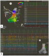
Three-dimensional left atrial electroanatomic mapping with an example of pre and post pulmonary veins isolation (respectively panel A and B) with a very high power-short duration strategy (CARTO System—90 watts for 4 seconds).
Figure 2
Figure 2
An example of three-dimensional electroanatomic mapping showing pre (panel A) and post (panel B) ablation with high power-short duration (ENSITE system—50 watts for 10 s).
Figure 3
Figure 3
A multimodal approach may contribute to detection of left atrial low voltage area. In these examples from the left to the right: voltage map during sinus rhythm (cutoff < 0.5 mV) and activation map performed with electroanatomical mapping (Ensite SJM), P wave duration, LA dimension detected by CT scan, and deformation detected by echo strain. On top, a case of normal voltage with normal conduction; the P wave is not prolonged, and deformation is normal in this case. In the middle, a case of prolonged P wave, LA enlargement at the CT scan, and impaired strain with moderate low voltage. On the bottom, severely dilated LA, P wave prolongation, and a severely impaired LA strain with a severe degree of low voltage area.
Figure 4
Figure 4
Pre-ablation voltage map of the left atrium with a posterior wall low voltage area (panel A). Post-processing CMR images 3D colorimetric map with ADAS 3D software (panel B). According to the software, the red areas relate to atrial fibrosis at the level of the posterior wall in this case, matching the low voltage area shown in panel A.
Figure 5
Figure 5
Recurrence of highly symptomatic paroxysmal AF after previous cryoablation (PVI alone) and empirical posterior wall isolation on a second procedure. At the time of the third procedure, the initial map shows persistent PVI and PWI (panel A). Drug challenging with high-dose isoproterenol is performed. Several multipolar catheters are placed in strategic positions to map extra PV triggers (in this case at the crista terminalis, SVC, LA anterior wall, and coronary sinus as shown in panel B). Biatrial ablation of extra PV (panel C). On panel D at the upper part, arrhythmia burden after the second procedure, on the bottom after extra PV trigger ablation as detected by implanted loop recorder.
Figure 6
Figure 6
Extra PV trigger elimination—During a de novo RF ablation procedure, spontaneous initiation of sustained AF arises from the superior vena cava. Circumferential SVC ablation results in sinus rhythm restoration (panel A), while AF persists confined to the SVC for the whole observation time (panel B).
Figure 7
Figure 7
Recurrence of symptomatic persistent atrial fibrillation after previous PVI with roof and anterior mitral lines. panel A On sinus rhythm, a three-dimensional electro-anatomical map of the left atrium shows persistent PV isolation and a low voltage (cutoff < 0.5 mV) area on the posterior and on the anterior LA wall. During repeated adenosine applications, evidence of left atrial appendage triggering atrial fibrillation (panel B). Linear ablation of the lateral mitral isthmus and conversion to sinus rhythm, while atrial fibrillation persists in the electrically isolated left atrial appendage (decapolar catheter). Modified from Madaffari A. et al. [81] Left Atrial Appendage Electrical Isolation for Persistent Atrial Fibrillation.
Figure 8
Figure 8
First redo ablation of persistent AF after PVI. The initial voltage map during AF shows severe low voltage with reconnection of the left pulmonary veins (upper panel). During VOM EI, LPV isolation occurs (Octaray catheter in the LSPV—lower panel).
Figure 9
Figure 9
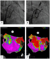
Vein of Marshall ethanol infusion for persistent atrial fibrillation ablation. Fluoroscopic images (right anterior oblique 30°) with VOM visualization after balloon inflation and contrast medium injection (panel A); staining lesion along the entire VOM course after ethanol infusion (panel B). 3D EAM from a posterior view with voltage map pre (panel C) and post VOMEI resulting in left inferior pulmonary vein isolation and partially posterior wall involvement (panel D).
Figure 10
Figure 10
PFA for atypical flutter ablation—Persistent flutter after PVI. A map of the ongoing flutter performed with a multipolar catheter (Octaray–CARTO) shows a scar-related atypical flutter localized to the LA posterior wall (panel A) left: activation map; right: voltage map during ongoing flutter). The Farapulse PFA catheter is visualized in this case as a circular catheter by the CARTO mapping system. A single PFA application at the critical isthmus, as shown, terminates the arrhythmia with restoration of sinus rhythm (panel B). Complete posterior wall isolation with additional application is successfully performed (panel C).

References

    1. Holmes D.N., Piccini J.P., Allen L.A., Fonarow G.C., Gersh B.J., Kowey P.R., O’Brien E.C., Reiffel J.A., Naccarelli G.V., Ezekowitz M.D., et al. Defining Clinically Important Difference in the Atrial Fibrillation Effect on Quality-of-Life Score. Circ. Cardiovasc. Qual. Outcomes. 2019;12:e005358. doi: 10.1161/CIRCOUTCOMES.118.005358. - DOI - PubMed
    1. Odutayo A., Wong C.X., Hsiao A.J., Hopewell S., Altman D.G., Emdin C.A. Atrial Fibrillation and Risks of Cardiovascular Disease, Renal Disease, and Death: Systematic Review and Meta-Analysis. BMJ. 2016;354:i4482. doi: 10.1136/bmj.i4482. - DOI - PubMed
    1. Bassand J.-P., Accetta G., Camm A.J., Cools F., Fitzmaurice D.A., Fox K.A.A., Goldhaber S.Z., Goto S., Haas S., Hacke W., et al. Two-Year Outcomes of Patients with Newly Diagnosed Atrial Fibrillation: Results from GARFIELD-AF. Eur. Heart J. 2016;37:2882–2889. doi: 10.1093/eurheartj/ehw233. - DOI - PMC - PubMed
    1. Kirchhof P., Camm A.J., Goette A., Brandes A., Eckardt L., Elvan A., Fetsch T., van Gelder I.C., Haase D., Haegeli L.M., et al. Early Rhythm-Control Therapy in Patients with Atrial Fibrillation. N. Engl. J. Med. 2020;383:1305–1316. doi: 10.1056/NEJMoa2019422. - DOI - PubMed
    1. Wilber D.J., Pappone C., Neuzil P., De Paola A., Marchlinski F., Natale A., Macle L., Daoud E.G., Calkins H., Hall B., et al. Comparison of Antiarrhythmic Drug Therapy and Radiofrequency Catheter Ablation in Patients with Paroxysmal Atrial Fibrillation. JAMA. 2010;303:333. doi: 10.1001/jama.2009.2029. - DOI - PubMed

LinkOut - more resources