. 2025 Mar 5;17(3):373.
doi: 10.3390/v17030373.

Oropouche Virus: Isolation and Ultrastructural Characterization from a Human Case Sample from Rio de Janeiro, Brazil, Using an In Vitro System

Affiliations

Oropouche Virus: Isolation and Ultrastructural Characterization from a Human Case Sample from Rio de Janeiro, Brazil, Using an In Vitro System

Ana Luisa Teixeira de Almeida et al. Viruses. .

Abstract

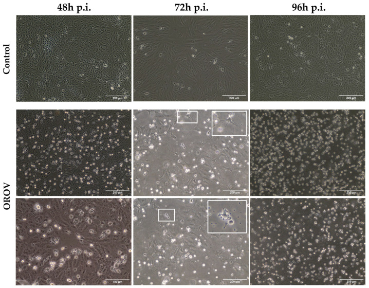
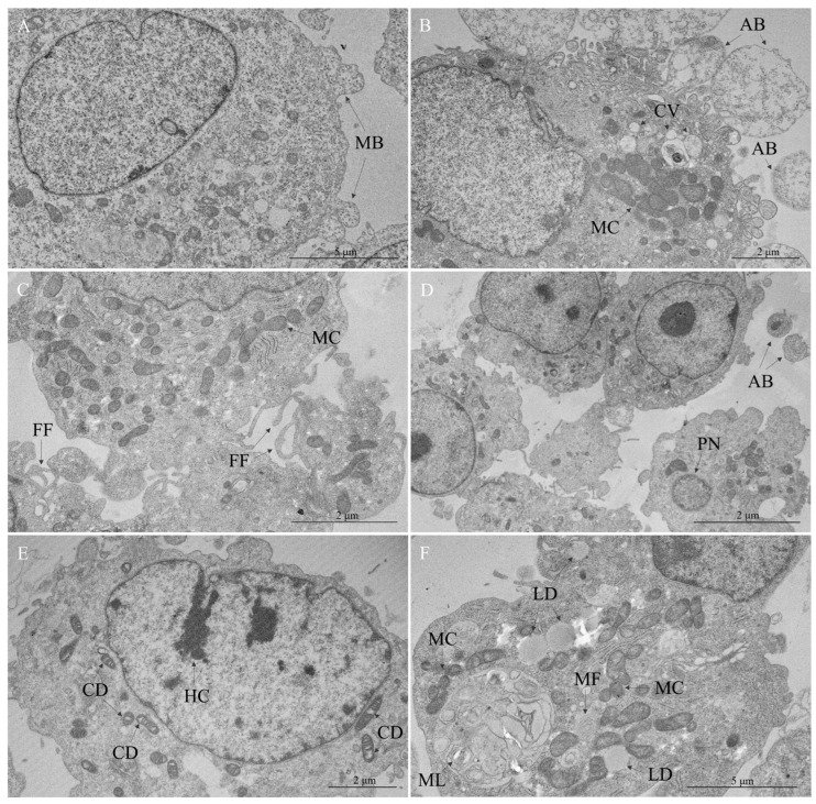
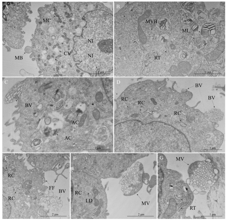
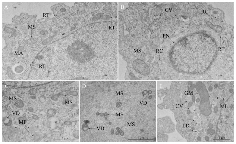
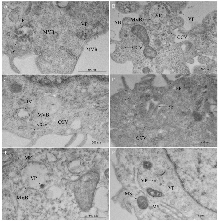
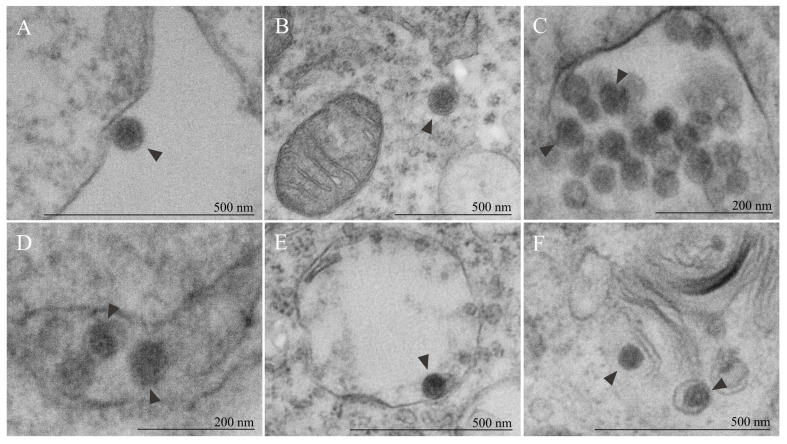
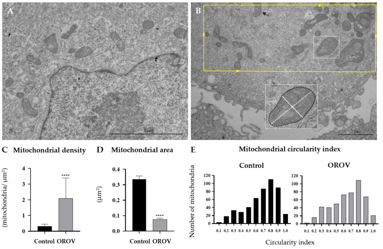
The Oropouche virus (OROV) is a segmented negative-sense RNA arbovirus member of the Peribunyaviridae family, associated with recurring epidemics of Oropouche fever in Central and South America. Since its identification in 1955, OROV has been responsible for outbreaks in both rural and urban areas, with transmission involving sylvatic and urban cycles. This study focuses on the characterization of an OROV isolate from a human clinical sample collected in the state of Rio de Janeiro, a non-endemic region in Brazil, highlighting ultrastructural and morphological aspects of the viral replicative cycle in Vero cells. OROV was isolated in Vero cell monolayers which, following viral inoculation, exhibited marked cytopathic effects (CPEs), mainly represented by changes in cell morphology, including membrane protrusions and vacuolization, as well as cell death. Studies by transmission electron microscopy (TEM) revealed significant ultrastructural changes, such as apoptosis, intense remodeling of membrane-bound organelles and signs of rough endoplasmic reticulum and mitochondrial stress. Additionally, the formation of specialized cytoplasmic vacuoles and intra- and extracellular vesicles emphasized trafficking and intercellular communication as essential mechanisms in OROV infection. RT-qPCR studies confirmed the production of viral progeny in high titers, corroborating the efficiency of this experimental model. These findings contribute to a better understanding of the cytopathogenic mechanisms of OROV infection and the contribution of cellular alterations in OROV morphogenesis.

Keywords: Oropouche virus; transmission electron microscopy; ultrastructural studies.


Conflict of interest statement

The authors declare no conflicts of interest.

Figures

Figure 1
Figure 1
Monolayers of Vero cells in control conditions and infected with Oropouche virus (OROV) at 48, 72, and 96 h post-infection (h p.i.). Bright-field microscopy analysis revealed conformational changes in the membrane as the infection progresses, including the formation of projections and membrane partitioning (insets), which are indicative of apoptosis.
Figure 2
Figure 2
Cytopathic effects in Vero cells infected with Oropouche virus—cellular response and damage. (A) Uninfected Vero cell. Membrane blebbing (MB). (BF) OROV-infected cells. (B) Apoptotic bodies (AB), cytoplasmic vacuoles (CV), mitochondrial clustering (MC). (C) Filopodia formation (FF), mitochondrial clustering (MC). (D) Apoptotic bodies (AB), pyknotic nucleus (PN). (E) Mitochondrial cristae disorganization (CD), hypercondensation of chromatin (HC). (F) Lipid droplets (LD), mitochondrial clustering (MC), myelin figure (MF), multilamellar bodies (ML).
Figure 3
Figure 3
Cytopathic effects in Vero cells infected with Oropouche virus—membrane remodeling. (A) Cytoplasmic vacuoles (CV), plasma membrane blebbing (MB), mitochondrial clustering (MC), nuclear membrane invagination (NI). (B) Multilamellar body (ML), multivesicular body (MVB), rough endoplasmic reticulum (RER) thickening (RT). (C) Autophagy components (AC), budding vesicles (BV). (D) Budding vesicles (BV), RER cisterns (RC). (E) Budding vesicles (BV), filopodia formation (FF), RER cisterns (RC). (F) Lipid droplet (LD), multivesicular cargo (MV), RER cisterns (RC). (G) Multivesicular cargo (MV), RER thickening (RT).
Figure 4
Figure 4
Cytopathic effects in Vero cells infected with Oropouche virus—rough endoplasmic reticulum (RER) and mitochondrial stress. (A) Mitophagy (MA), mitochondrial swelling (MS), RER thickening (RT). (B) Cytoplasmic vacuole (CV), mitochondrial swelling (MS), pyknotic nucleus (PN), RER cisterns (RC), RER thickening (RT). (C) Myelin figures (MF), mitochondrial swelling (MS), vacuolar degeneration of mitochondria (VD). (D) Mitochondrial swelling (MS), vacuolar degeneration of mitochondria (VD). (E) Cytoplasmic vacuole (CV), giant mitochondria (GM), lipid droplets (LD), multilamellar bodies (ML).
Figure 5
Figure 5
Cytopathic effects in Vero cells infected with Oropouche virus—viral replication insights. (A,B) Multivesicular body (MVB), viral particles (VP). (A) Immature viral particles (IP), intraluminal vesicle (IV). (BD) Clathrin-coated vesicles (CCV). (B) Apoptotic body (AB). (C) Intraluminal vesicle (IV), multivesicular body (MVB). (D) Filopodia formation (FF). (E,F) Viral particles (VP). (E) Multivesicular body (MVB), myelin figures (MF). (F) Mitochondrial swelling (MS).
Figure 6
Figure 6
Morphology of Oropouche viral particles. (A) Viral particle (arrowhead) in extracellular space, adsorbed to the membrane surface. (BF) Viral particles (arrowhead) into cytoplasm. (CE) Morphological diversity noted within multivesicular bodies, showcasing particles at different stages of morphogenesis (arrowhead), leading to variations in size and electron density. (C) Reduced diameter observed in indicated particles (arrowhead) reflects incomplete particles. (F) Additional location of viral particles (arrowhead) within cytoplasm, associated with Golgi apparatus stacks. Inset: (C,D) Figure 5A. (E) Figure 5E. (F) Figure 5F. All particles exhibited a spherical shape, with variation in diameter indirectly reflecting maturation status of these particles.
Figure 7
Figure 7
Morphometric analysis of the mitochondria. (A) Micrograph of the uninfected cell. (B) Micrograph of the OROV-infected cell culture to illustrate morphometric procedure. All mitochondria were counted, excluding those that were not fully within the field (black arrow). For the mitochondrial density of each micrography, the evaluated areas (yellow rectangle) were defined as the boundary surrounding the outermost mitochondria (yellow crosses). The cross-sectional area (black dashed line) and minor and major axes (continuous and dashed white line, respectively) were measured in all mitochondria (C) Mitochondrial density of 20 micrographs per group. Median: 0.29 mitochondria/μm2 (95% CI, 0.25–0.31)—control group), and 1.78 mitochondria/μm2 (95% CI, 1.27–2.3)—OROV-infected group. The data presented in panels (C,D) correspond to the micrographs totaling 500 mitochondria per group (control and OROV-infected). (D) Mitochondrial cross-sectional area. Median: 0.37 μm2 (95% CI, 0.32–0.36)—control group, and 0.08 μm2 (95% CI, 0.07–0.08)—OROV-infected group. **** p < 0.0001—Mann–Whitney test. (E) Distribution of the frequencies of mitochondrial circularity index. The y-axis indicates the number of mitochondria located within the circularity index range specified by the center bins on the x-axis. Median: 0.72—control group, and 0.68—OROV-infected group.

Similar articles

References

    1. Anderson C.R., Spence L., Downs W.G., Aitken T.H. Oropouche virus: A new human disease agent from Trinidad, West Indies. Am. J. Trop. Med. Hyg. 1961;10:574–578. doi: 10.4269/ajtmh.1961.10.574. - DOI - PubMed
    1. Sakkas H., Bozidis P., Franks A., Papadopoulou C. Oropouche Fever: A Review. Viruses. 2018;10:175. doi: 10.3390/v10040175. - DOI - PMC - PubMed
    1. Pinheiro F., Pinheiro M., Bensabath G., Causey O.R., Shope R.E. Epidemia de vírus Oropouche em Belém. Rev. Serv. Espec. Saude Publica. 1962;12:13–23.
    1. Sah R., Srivastava S., Kumar S., Golmei P., Rahaman S.A., Mehta R., Ferraz C., Apostolopoulos V., Rodriguez-Morales A.J. Oropouche fever outbreak in Brazil: An emerging concern in Latin America. Lancet Microbe. 2024;5:100904. doi: 10.1016/S2666-5247(24)00136-8. - DOI - PubMed
    1. Azevedo R.S., Nunes M.R., Chiang J.O., Bensabath G., Vasconcelos H.B., Pinto A.Y., Martins L.C., Monteiro H.A., Rodrigues S.G., Vasconcelos P.F. Reemergence of Oropouche fever, northern Brazil. Emerg. Infect. Dis. 2007;13:912–915. doi: 10.3201/eid1306.061114. - DOI - PMC - PubMed

Publication types

Supplementary concepts

LinkOut - more resources