. 2025 Mar 12;17(3):401.
doi: 10.3390/v17030401.

The Infectivity and Pathogenicity Characteristics of a Recombinant Porcine Epidemic Diarrhea Virus, CHFJFQ

Affiliations

The Infectivity and Pathogenicity Characteristics of a Recombinant Porcine Epidemic Diarrhea Virus, CHFJFQ

Zhihua Feng et al. Viruses. .

Abstract

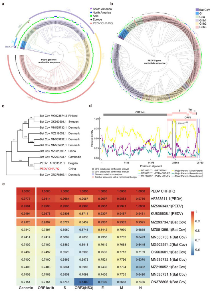
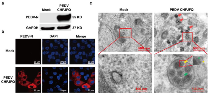
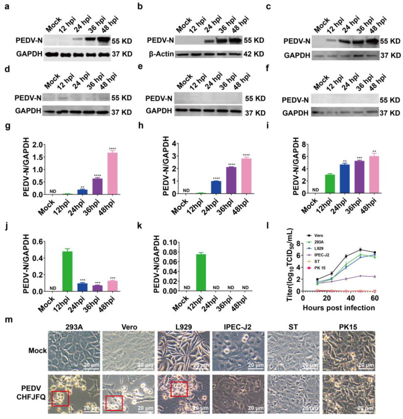
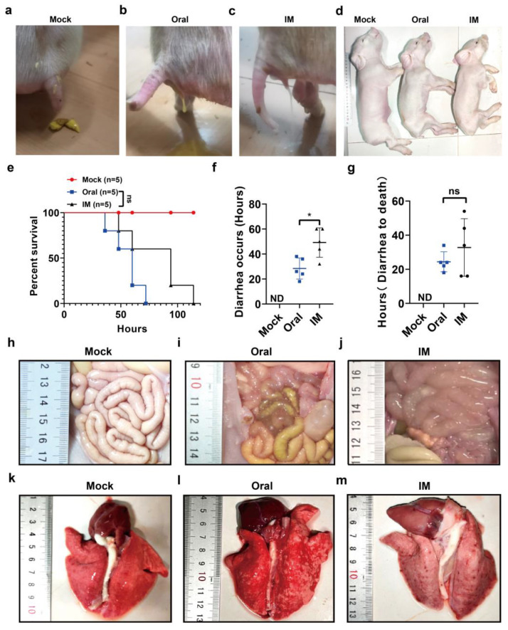
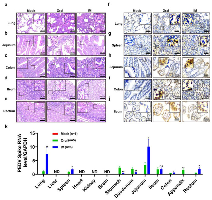
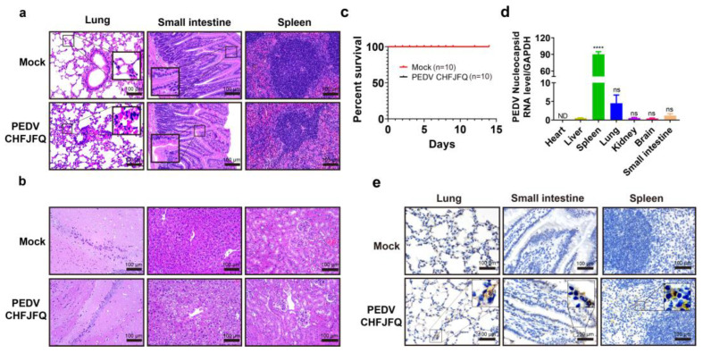
Porcine epidemic diarrhea virus (PEDV) presents a substantial challenge to the global swine industry. However, the origin, host range, and potential cross-species transmission of PEDV remain poorly understood. This study characterizes a novel PEDV strain, CHFJFQ, isolated from diarrheic piglets in Fuqing, Fujian, China. Through sequencing and phylogenetic analysis, we determined that CHFJFQ belongs to the GIIa subgroup and is a recombinant with CH/HNXX/2016 as the major parent and NW17 as the minor parent. Compared to CV777, CHFJFQ exhibits multiple base deletions and insertions across the 5'UTR, ORF1a/b, S, and ORF3 genes. Phylogenetic analysis indicates shared ancestry with bat coronaviruses, though a direct zoonotic origin remains uncertain. Interestingly, CHFJFQ demonstrated its ability to infect human and mouse cell lines in vitro and, more significantly, caused in vivo infection in both pigs and mice. The primary target organs were the intestines, lungs, and spleen, resulting in 100% mortality in suckling piglets. PEDV CHFJFQ was detected in mouse tissues, but no clinical signs were observed, indicating limited cross-species pathogenicity. Overall, these findings offer crucial insights into the epidemiology, genetics, infectivity, and pathogenicity of PEDV and provide valuable information for vaccine development.

Keywords: PEDV; cross-species transmission; infection; origin.


Conflict of interest statement

The authors declare no conflicts of interest.

Figures

Figure 1
Figure 1
Nucleotide deletions and insertions in the genome of PEDV CHFJFQ compared to CV777, AJ1102, and DR13. (a) Blue strip: Polyprotein 1a and non-structural proteins 1–11; red strip: polyprotein 1b and non-structural proteins 12–16; green strip: spike protein (S); brown: ORF3; yellow strip: envelope protein; purple strip: membrane protein; orange strip: nucleocapsid protein. (b) Compared to the CV777 strain, 5 nucleotides were deleted, and 1 nucleotide was inserted in the 5′ UTR of CHFJFQ, AJ1102, virulent DR13, and attenuated DR13. (c) Compared to the CV777 strain, a 24-nucleotide deletion was observed in the ORF1a/b region of both the CHFJFQ and attenuated DR13 strains. (d) Compared to the CV777 strain, CHFJFQ and attenuated DR13 strains showed a 3-nucleotide deletion in the S gene, while AJ1102 exhibited a 6-nucleotide deletion and 3-nucleotide insertion in the S gene. (e) Based on the CV777 strain, a 49-nucleotide deletion was observed in the ORF3 region of both CHFJFQ and attenuated DR13. (f) Compared to CV777, CHFJFQ, AJ1102, and virulent DR13 strains, a 21-nucleotide deletion was observed in the E gene of the attenuated DR13 strain. (g,h) No nucleotide deletions or insertions were observed in the M or N gene of the CV777, CHFJFQ, AJ1102, virulent DR13, and attenuated DR13 strains. Sequence alignment was performed using BioEdit software (Version 7.0.9.0), with ClustalW multiple alignment selected (Full Multiple Alignment, Bootstrap NJ Tree, number of bootstraps set to 1000).
Figure 2
Figure 2
Phylogenetic analysis, nucleotide similarity, and recombination analysis of PEDV CHFJFQ. To explore the evolution and recombination of PEDV, 245 complete PEDV genome sequences and 2 bat coronavirus genome sequences were analyzed using MUSCLE, IQtree, MEGA7, and RDP5. PEDV CHFJFQ, marked with red stars, belongs to the GIIa subgroup. (a) The evolution and global distribution of PEDV were analyzed based on genome-wide data. (b) The evolution and global distribution of PEDV were also analyzed based on the S gene. (c) Phylogenetic analyses of PEDVs and bat coronaviruses. (d) PEDV CHFJFQ is potentially a recombinant strain, with CH/HNXX/2016 as the major parent and NW17 as the minor parent. The recombination regions are highlighted in pink. (e) Nucleotide similarity analyses of PEDV and bat coronavirus genomes. MUSCLE software was used to align 247 full-length genome sequences, including 245 PEDV and 2 bat coronavirus genomes. The following command was executed: muscle -in seqs.fa -out seqs.afa -maxiters 2, with a maximum iteration limit of 2. Subsequently, IQtree software was used to construct rootless phylogenetic trees based on a maximum likelihood algorithm, executed with the command iqtree -s seqs.afa -nt 50 -iteration 2. ModelFinder was used to assess various nucleotide substitution models and select the most suitable one for constructing the evolutionary tree. The phylogenetic tree data were further analyzed using the interactive Tree Of Life (iTOL) platform (https://itol.embl.de/, accessed on 12 February 2025). Additionally, Recombination Detection Program version 5 (RDP5) was used to identify potential recombination events within the PEDV CHFJFQ strain. For the phylogenetic tree analysis of the S genes of 9 bat coronaviruses and 2 PEDV strains (PEDV CHFJFQ and PEDV CV777), MEGA software was used. Sequence alignment across 11 groups was performed using MUSCLE integrated within MEGA, producing 11 sets of aligned S protein sequences. A phylogenetic tree was then constructed using a maximum likelihood approach. BLAST was applied to compare different gene positions across multiple groups, providing similarity values for each gene. The resulting data were visualized as a heatmap using the R package pheatmap.
Figure 3
Figure 3
The expression of PEDV N protein and virus particles in Vero cells. (a) The expression of PEDV N protein in Vero cells was detected by Western blotting at 48 hpi. Vero cells (5 million per well) in six-well plates were co-cultured with PEDV CHFJFQ (MOI: 0.1). Cells were harvested at 48 hpi, and total protein was extracted for Western blotting. Mouse monoclonal anti-PEDV N protein antibody was used to detect the expression of PEDV N protein, with GAPDH as the control. (b) The expression of PEDV N protein in Vero cells was detected by immunofluorescence at 48 hpi. Vero cells (1 × 10⁵) were co-cultured with PEDV CHFJFQ (MOI = 0.1) in confocal dishes. Cells were fixed, permeabilized, and probed with mouse monoclonal anti-PEDV N protein antibody, followed by Alexa Fluor™ 546 Donkey anti-mouse IgG (H + L) (red) for confocal immunofluorescence detection of PEDV N protein expression. Nuclei were stained with DAPI (blue). (c) Morphology of PEDV virions observed by transmission electron microscopy (48 hpi). Vero cells (5 × 10⁵ per well) were co-cultured with PEDV CHFJFQ (MOI: 0.01) in six-well plates. Cells were fixed at 48 hpi for subsequent TEM examination. Virus particles (red arrow): 80–100 nm; envelope (green arrow): 5–7 nm; spike (yellow arrow): 16–20 nm. The experiment was independently repeated three times, and the presented figure depicts the results of one representative replicate. Full-length blots are presented in Supplementary Figure S4.
Figure 4
Figure 4
Proliferative characteristics of PEDV CHFJFQ in vitro. (af) The expression of PEDV N protein in 293A, Vero, L929, IPEC-J2, ST, and PK15 cells was detected by Western blotting at various time points post-infection (n = 3). Six cell lines (5 × 10⁵ cells per well) were co-cultured with PEDV CHFJFQ (MOI = 0.1) in 6-well plates. Cells were harvested at 12, 24, 36, and 48 h post-infection (hpi), and total protein was extracted for Western blot analysis. Anti-PEDV N protein antibody was used to detect PEDV N protein expression, and GAPDH or β-actin was used as endogenous controls. (gk) The relative expression of PEDV N protein in relation to GAPDH or β-actin from (ae) was analyzed using ImageJ (v.1.8.0) (n = 3). (l) Viral titers at different time points of PEDV infection in 293A, Vero, L929, IPEC-J2, ST, and PK15 cells. Six cell lines (5 × 10⁵ cells per well) were co-cultured with PEDV CHFJFQ (MOI = 0.1) in 6-well plates. The culture supernatants were harvested at 12, 24, 36, and 48 hpi, and the virus titers were determined using the Spearman–Kärber method (n = 3). (m) Cytopathic effects were observed by microscopy and photographed at 48 hpi of PEDV CHFJFQ infection. The red box highlights the cytopathic effects caused by PEDV CHFJFQ infection. Unpaired t-tests (GraphPad Prism 5.0, GraphPad Software, San Diego, CA, USA) were used to test differences between groups. Data are presented as means ± standard error of the mean for each treatment. ** p < 0.01, *** p < 0.001, and **** p < 0.0001 vs. control group (12 hpi); ND: none detected. Sample sizes are indicated in brackets. The full-length blots are presented in Supplementary Figure S7.
Figure 5
Figure 5
Clinical symptoms and survival of piglets challenged with PEDV CHFJFQ. (ac) Both oral and intramuscular inoculation of PEDV CHFJFQ resulted in diarrhea. (d) Dehydration was observed in piglets following both oral and intramuscular inoculation of PEDV CHFJFQ (60 hpi). (e) Survival curves of piglets challenged with PEDV CHFJFQ (n = 5). (f) Time to onset of diarrhea following inoculation with PEDV CHFJFQ (n = 5). (g) Time to death in piglets due to diarrhea (n = 5). (hj) Oral or intramuscular inoculation of PEDV CHFJFQ caused intestinal thinning in piglets. (km) Pulmonary hemorrhage was observed in piglets following oral or intramuscular inoculation with PEDV CHFJFQ. The control group was inoculated with 1 mL of DMEM via oral and intramuscular injection, respectively. The oral and intramuscular inoculation groups received 1 mL of 5 × 10⁶ TCID50 PEDV CHFJFQ. The health status of the piglets was monitored every 3 h, and the experiment was terminated when piglets were unable to stand or eat. Data were analyzed using the Mantel–Cox test (e), and unpaired t-tests (f,g) (GraphPad Prism 5.0, GraphPad Software, San Diego, CA, USA) were used to test differences between groups. Data are presented as means ± standard error of the mean for each treatment. * p < 0.05 vs. control group (oral), ns: p > 0.05 vs. control group (oral), ND: none detected. Sample sizes are indicated in brackets.
Figure 6
Figure 6
Pathological changes induced by PEDV CHFJFQ and its distribution in piglets. (a) PEDV CHFJFQ causes thickening of the alveolar walls and pulmonary inflammation in piglets. (b) PEDV CHFJFQ induces villous atrophy and epithelial cell shedding in the jejunum of piglets. (c) The colon of a piglet inoculated with PEDV CHFJFQ shows diffuse atrophic enteritis. (d) PEDV CHFJFQ leads to the shedding of villous epithelial cells in the ileum of piglets. (e) Intramuscular inoculation of PEDV CHFJFQ induces shedding of rectal villus epithelial cells in piglets. (fj) Immunohistochemical analysis of PEDV N protein expression in the lung, spleen, jejunum, ileum, and colon. (k) The relative content of the PEDV CHFJFQ S gene in each organ was detected by RT-qPCR (n = 5). ND: none detected. Unpaired t-tests (GraphPad Prism 5.0, GraphPad Software, San Diego, CA, USA) were used to test differences between groups. Data are presented as means ± standard error of the mean for each treatment. * p < 0.05, ** p < 0.01 vs. control group (oral), ns: p > 0.05 vs. control group (oral), ND: none detected. Sample sizes are indicated in brackets.
Figure 7
Figure 7
Clinical symptoms, survival curves, and pathological changes in mice challenged with PEDV CHFJFQ. (a,b) PEDV CHFJFQ caused lung and liver injury as well as intestinal villus atrophy in mice. Hematoxylin–eosin (H&E) staining was used to observe the pathological changes in mice induced by PEDV CHFJFQ infection at 72 hpi (n = 5). (c) Survival curves of mice challenged with PEDV CHFJFQ (n = 10). (d) The relative content of the PEDV CHFJFQ N gene in each organ was detected by RT-qPCR (n = 5). (e) Immunohistochemical detection of PEDV N protein expression in the lung, jejunum, and spleen at 72 hpi (n = 5). Mice in the PEDV infection group were orally inoculated with 5 × 10⁵ TCID50 of PEDV CHFJFQ (0.1 mL), while the control group received the same volume of DMEM. Unpaired t-tests (GraphPad Prism 5.0, GraphPad Software, San Diego, CA, USA) were used to test differences between groups. Data are presented as means ± standard error of the mean for each treatment. **** p < 0.0001 vs. control group (liver), ns: p > 0.05 vs. control group (liver), ND: none detected. Sample sizes are indicated in brackets.

Similar articles

References

    1. Li Z., Ma Z., Dong L., Yang T., Li Y., Jiao D., Han W., Zheng H., Xiao S. Molecular Mechanism of Porcine Epidemic Diarrhea Virus Cell Tropism. mBio. 2022;13:e0373921. doi: 10.1128/mbio.03739-21. - DOI - PMC - PubMed
    1. Brian D.A., Baric R.S. Coronavirus genome structure and replication. Curr. Top. Microbiol. Immunol. 2005;287:1–30. - PMC - PubMed
    1. Niu X., Kong F., Hou Y.J., Wang Q. Crucial mutation in the exoribonuclease domain of nsp14 of PEDV leads to high genetic instability during viral replication. Cell Biosci. 2021;11:106. doi: 10.1186/s13578-021-00598-1. - DOI - PMC - PubMed
    1. Wang D., Ge X., Chen D., Li J., Cai Y., Deng J., Zhou L., Guo X., Han J., Yang H. The S Gene Is Necessary but Not Sufficient for the Virulence of Porcine Epidemic Diarrhea Virus Novel Variant Strain BJ2011C. J. Virol. 2018;92:e00603-18. doi: 10.1128/JVI.00603-18. - DOI - PMC - PubMed
    1. Li Z., Ma Z., Li Y., Gao S., Xiao S. Porcine epidemic diarrhea virus: Molecular mechanisms of attenuation and vaccines. Microb. Pathog. 2020;149:104553. doi: 10.1016/j.micpath.2020.104553. - DOI - PMC - PubMed

Publication types

Associated data

LinkOut - more resources