An Aberrant Right Subclavian Artery Causing Severe Esophageal Compression: A Case Report
- PMID: 40144436
- PMCID: PMC11937618
- DOI: 10.7759/cureus.79527
An Aberrant Right Subclavian Artery Causing Severe Esophageal Compression: A Case Report
Abstract
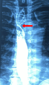
An aberrant right subclavian artery (ARSA), a congenital vascular anomaly, can cause significant esophageal compression, leading to a condition known as dysphagia lusoria (DL). We present the case of a 44-year-old man with progressively worsening dysphagia and odynophagia over the last six months, resulting in severe weight loss and dietary restrictions. Imaging techniques revealed esophageal stenosis caused by external compression from an ARSA arising from the posterior wall of the distal aortic arch, accompanied by a Kommerell's diverticulum. Computed tomography angiography confirmed the aberrant origin, retroesophageal course, and vascular anomaly. Although surgical intervention involving ligation and excision of the retroesophageal artery segment with a right carotid-subclavian bypass was recommended, the patient opted for conservative management. This case highlights the importance of advanced imaging techniques in diagnosing DL and guiding treatment decisions. Regular follow-up remains essential to monitor disease progression and manage potential complications.
Keywords: aberrant right subclavian artery; dysphagia lusoria; esophageal compression; kommerell’s diverticulum; vascular anomalies.
Copyright © 2025, Galanis et al.
Conflict of interest statement
Human subjects: Consent for treatment and open access publication was obtained or waived by all participants in this study. Conflicts of interest: In compliance with the ICMJE uniform disclosure form, all authors declare the following: Payment/services info: All authors have declared that no financial support was received from any organization for the submitted work. Financial relationships: All authors have declared that they have no financial relationships at present or within the previous three years with any organizations that might have an interest in the submitted work. Other relationships: All authors have declared that there are no other relationships or activities that could appear to have influenced the submitted work.
Figures


References
-
- Standring S. Edinburgh: Elsevier Churchill Livingstone; 2016. Gray's anatomy.
-
- Netter F. Philadelphia: Saunders; 2019. Atlas of human anatomy.
-
- Moore KL, Dalley AF, Agur AMR. Philadelphia: Lippincott Williams & Wilkins; 2014. Clinically oriented anatomy.
-
- Intermittent esophageal dysphagia: an intriguing diagnosis. Dysphagia lusoria. Gnanapandithan K, Rahni DO, Habr F. Gastroenterology. 2014;146:0–4. - PubMed
-
- Demonstration of vascular abnormalities compressing esophagus by MDCT: special focus on dysphagia lusoria. Alper F, Akgun M, Kantarci M, et al. Eur J Radiol. 2006;59:82–87. - PubMed
Publication types
LinkOut - more resources
Full Text Sources
