. 2024 Aug 12;14(1):36-57.
doi: 10.1159/000540180. eCollection 2025 Mar.

The Impact of TP53-Induced Glycolysis and Apoptosis Regulator on Prognosis in Hepatocellular Carcinoma: Association with Tumor Microenvironment and Ferroptosis

Affiliations

The Impact of TP53-Induced Glycolysis and Apoptosis Regulator on Prognosis in Hepatocellular Carcinoma: Association with Tumor Microenvironment and Ferroptosis

Katsuya Toshida et al. Liver Cancer. .

Abstract

Introduction: TP53-induced glycolysis and apoptosis regulator (TIGAR) is a p53 target protein that has critical roles in glycolysis and redox balance. The reports about the effect of TIGAR on prognosis and its biological role in hepatocellular carcinoma (HCC) are limited.

Methods: A total of 386 patients with HCC who had undergone hepatic resection were enrolled. Immunohistochemical staining for TIGAR was performed. Additionally, the regulation of malignant activity and ferroptosis by TIGAR was investigated in vitro.

Results: Patients were divided into TIGAR-positive (n = 80, 20.7%) and -negative (n = 306, 79.3%) groups. TIGAR positivity was significantly correlated with lower albumin, higher α-fetoprotein/ des-gamma-carboxyprothrombin, larger tumor size/number of tumors, and greater proportions of BCLC staging C/single nodular type/poor differentiation/microscopic vascular invasion/microscopic intrahepatic metastasis. In multivariate analysis, TIGAR positivity was an independent prognostic factor (p < 0.0001). In addition, TIGAR positivity was significantly associated with a smaller number of cluster of differentiation (CD) 8-positive T cells (p = 0.0450), larger number of CD68-positive macrophages (p = 0.0058), larger number of programmed death-ligand 1-positive cases (p = 0.0002), and larger number of vessels that encapsulate tumor cluster-positive cases (p = 0.0004). In vitro, TIGAR knockdown decreased cell motility and induced ferroptosis. TIGAR knockdown inhibited the phosphorylation of adenosine monophosphate-activated protein kinase and acetyl-CoA carboxylase. Ferroptosis induced by TIGAR knockdown was inhibited by liproxstatin and baicalein treatment. The combination of TIGAR knockdown and lenvatinib further induced ferroptosis.

Conclusion: High expression of TIGAR impacted the clinical outcome of HCC patients and TIGAR was associated not only with tumor microenvironment but also with resistance to ferroptosis.

Keywords: Ferroptosis; Hepatocellular carcinoma; Lenvatinib; TIGAR.


Conflict of interest statement

The authors have no conflict of interest.

Figures
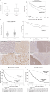
Fig. 1.
Fig. 1.
TIGAR is an indicator of poor prognosis in a public dataset and our cohort. aTIGAR mRNA expression level in tumor tissue compared with normal tissue using the TCGA dataset. OS in patients with HCC according to TIGAR mRNA expression. bTIGAR mRNA expression levels in tumor tissue compared with normal tissue using The International Cancer Genome Consortium dataset. OS in patients with HCC according to TIGAR mRNA expression. c IHC of TIGAR in patients with HCC. d IHC of TIGAR in cancerous and adjacent non-cancerous areas. e DFS in HCC patients according to TIGAR expression in our cohort. f OS in HCC patients according to TIGAR expression in our cohort. DFS, disease-free survival; HCC, hepatocellular carcinoma; OS, overall survival; TCGA, The Cancer Genome Atlas; TIGAR, TP53-induced glycolysis and apoptosis regulator.
Fig. 2.
Fig. 2.
TIGAR promotes HCC cell motility. Expression (a) and TIGAR knockdown (b) in HCC cell lines. Knockdown (KD) of TIGAR significantly inhibited cell viability (c), invasion (d), migration (e), and colony formation (f) in HCC cell lines compared with the negative control (Ctrl). g Gene set enrichment analysis (GSEA) using public dataset in HCC: biological process with significant difference between the high- and low-TIGAR expression groups, VEGF-related enrichment plots: VEGF production (NES: 1.76, FDR <0.0001), cellular response to VEGF stimulus (NES: 1.60, FDR <0.0001), and VEFG signaling pathway (NES: 1.58, FDR <0.0001). h TIGAR knockdown decreased the expression of ERK/phospho-ERK. d Scale bar: 200 μm. e Migration index: 1−[length at 24 h]/[length at 0 h], *p < 0.05, **p < 0.001, ***p < 0.0001. Ctrl, negative control; FDR, false discovery rate; HCC, hepatocellular carcinoma; KD, knockdown; NES, normalized enrichment score; TIGAR, TP53-induced glycolysis and apoptosis regulator; VEGF, vascular endothelial growth factor.
Fig. 3.
Fig. 3.
The association between TIGAR and tumor immune microenvironment. a IHC of CD8-low/high in patients with HCC. The number of CD8-positive T cells in TIGAR-negative/positive group. b IHC of CD68-low-high in patients with HCC. The number of CD68-positive macrophages in TIGAR-negative/positive group. c IHC of PD-L1-negative/positive in patients with HCC. The number of PD-L1 positive cases in TIGAR-negative/positive group. d IHC of VETC-negative/positive in patients with HCC. The number of VECT-positive cases in TIGAR-negative/positive group. CD, cluster of differentiation; HCC, hepatocellular carcinoma; PD-L1, programmed death-ligand 1; TIGAR, TP53-induced glycolysis and apoptosis regulator, VETC, vessels that encapsulate tumor cluster.
Fig. 4.
Fig. 4.
TIGAR knockdown increases ROS and promotes lipid peroxidation by inhibiting the AMPK-ACC pathway. TIGAR KD increased intracellular ROS levels (a), promoted lipid peroxidation (MDA levels) (b), and inhibited phosphorylation of AMPK and ACC in HCC cell lines compared with the Ctrl (c). Compound C (10 μm) inhibited the phosphorylation of AMPK and ACC (d) and promoted lipid peroxidation (e) in HCC cell lines. Induction of ferroptosis by TIGAR KD was significantly suppressed by10 μm LS (f) and 30 μm BC (g). TIGAR KD did not change the caspase-3/7 activity (relative fluorescent unit) in Huh7, Hep3B, and PLC (h). *p < 0.05, **p < 0.001, ***p < 0.0001. ACC, acetyl-CoA carboxylase; AMPK, adenosine monophosphate-activated protein kinase; BC, baicalein; Ctrl, negative control; HCC, hepatocellular carcinoma; KD, knockdown; LS, liproxstatin; MDA, malondialdehyde; TIGAR, TP53-induced glycolysis and apoptosis regulator.
Fig. 5.
Fig. 5.
Induction of ferroptosis by TIGAR knockdown in combination with lenvatinib (LEN). The combination of TIGAR KD and 10 μm LEN significantly decreased cell viability (a) and increased MDA levels (b) compared with knockdown + DMSO and Ctrl + LEN. *p < 0.05, **p < 0.001, ***p < 0.0001. Ctrl, negative control; LEN, lenvatinib; MDA, malondialdehyde; NC, negative control; TIGAR, TP53-induced glycolysis and apoptosis regulator.

References

    1. Global Burden of Disease Cancer Collaboration; Fitzmaurice C, Abate D, Abbasi N, Abbastabar H, Abd-Allah F, et al. . Global, regional, and national cancer incidence, mortality, years of life lost, years lived with disability, and disability-adjusted life-years for 29 cancer groups, 1990 to 2017: a systematic analysis for the global burden of disease study. JAMA Oncol. 2019;5(12):1749–68. - PMC - PubMed
    1. Okamura Y, Sugiura T, Ito T, Yamamoto Y, Ashida R, Ohgi K, et al. . Changes in patient background and prognosis after hepatectomy for hepatocellular carcinoma by hepatitis virus infection status: new trends in Japan. Ann Gastroenterol Surg. 2021;5(4):553–66. - PMC - PubMed
    1. Forner A, Reig M, Bruix J. Hepatocellular carcinoma. Lancet. 2018;391(10127):1301–14. - PubMed
    1. Itoh S, Yugawa K, Shimokawa M, Yoshiya S, Mano Y, Takeishi K, et al. . Prognostic significance of inflammatory biomarkers in hepatocellular carcinoma following hepatic resection. BJS Open. 2019;3(4):500–8. - PMC - PubMed
    1. Iseda N, Itoh S, Yoshizumi T, Tomiyama T, Morinaga A, Shimagaki T, et al. . Lymphocyte-to-C-reactive protein ratio as a prognostic factor for hepatocellular carcinoma. Int J Clin Oncol. 2021;26(10):1890–900. - PubMed