

Sagittal changes in the dens significantly slowed after 12 years of age
- PMID: 40144789
- PMCID: PMC11938264
- DOI: 10.1016/j.bas.2025.104233
Sagittal changes in the dens significantly slowed after 12 years of age
Abstract
Introduction: The odontoid process is an important component of the upper cervical, and the process of ossification for odontoid does not cease completely until skeletal maturity.
Research question: The aim of the study was to obtain the inclination parameters of the dens in many healthy children and to analyze the trends and variations in the inclination of the dens.
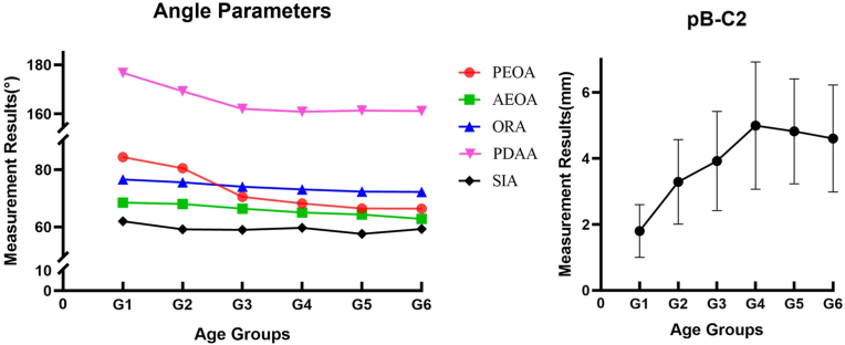
Material and methods: All the CT data obtained from our hospital was reviewed for next measurement. Posterior edge of odontoid angle (PEOA), anterior edge of odontoid angle (AEOA), odontoid retroflection angle (ORA), posterior dens angulation angle (PDAA), screw insertion angle (SIA), and pB-C2 line were measured and analyzed.
Results: A total of 219 patients were divided into 6 groups based on age at an interval of 3 years. The mean values of PEOA and PDAA dropped dramatically with age up to the ten to twelve-year group and then decreased slightly until 18 years old. Moreover, the AEOA and ORA declined gradually from birth to adulthood. These parameters were statistical significance within different age groups. However, the SIA was largely unchanged from birth to 18 years old and appeared to be independent of age. In contrast, the PB-C2 line has a distinguish distribution, with an increase up to the nine to twelve-year age group and then gradually decreased until 18 years old.
Discussion and conclusion: The inclination of dens was constantly changing during pediatric growth, but the trends were different. These developmental changes slow down significantly after the age of 12 years.
Keywords: Cervical spine; Growth; Inclination; Odontoid; Pediatric.
© 2025 The Authors.
Conflict of interest statement
The authors declare that they have no known competing financial interests or personal relationships that could have appeared to influence the work reported in this paper.
Figures

References
-
- Baaghtmyr El-Hawary. In: The Growing Spine. third ed. Alain Dimeglio F.C., Bonnel François, Parent Stefan, editors. Springer Nature; Switzerland: 2022. p. 58.
-
- Chauhan A.K., Chandra P.S., Goyal N., Chowdhury M.R., Banerjee J., Tripathi M., et al. Weak ligaments and sloping joints: a new hypothesis for development of congenital atlantoaxial dislocation and basilar invagination. Neurospine. 2020;17(4):843–856. doi: 10.14245/ns.2040434.217. Epub 2021/01/07. PubMed PMID: 33401861; PubMed Central PMCID: PMCPMC7788422. - DOI - PMC - PubMed
-
- Chun D.H., Yoon D.H., Kim K.N., Yi S., Shin D.A., Ha Y. Biomechanical Comparison of four different atlantoaxial posterior fixation constructs in adults: a finite element study. Spine (Phila Pa 1976) 2018;43(15) doi: 10.1097/brs.0000000000002584. E891-e7. Epub 2018/02/21. PubMed PMID: 29462065. - DOI - PubMed
LinkOut - more resources
Full Text Sources
Miscellaneous
