. 2025 Mar 12:16:1532852.
doi: 10.3389/fimmu.2025.1532852. eCollection 2025.

Guselkumab binding to CD64+ IL-23-producing myeloid cells enhances potency for neutralizing IL-23 signaling

Affiliations

Guselkumab binding to CD64+ IL-23-producing myeloid cells enhances potency for neutralizing IL-23 signaling

Kacey L Sachen et al. Front Immunol. .

Abstract

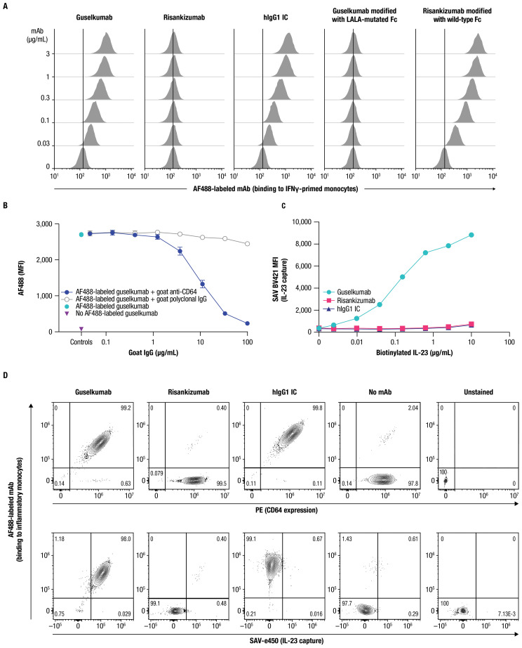
IL-23 is implicated in the pathogenesis of immune-mediated inflammatory diseases, and myeloid cells that express Fc gamma receptor 1 (FcγRI or CD64) on their surface have been recently identified as a primary source of IL-23 in inflamed tissue. Our complementary analyses of transcriptomic datasets from psoriasis and IBD showed increased expression of CD64 and IL-23 transcripts in inflamed tissue, and greater abundance of cell types with co-expression of CD64 and IL-23. These findings led us to explore potential implications of CD64 binding on the function of IL-23-targeting monoclonal antibodies (mAbs). Guselkumab and risankizumab are mAbs that target the IL-23p19 subunit. Guselkumab has a native Fc domain while risankizumab contains mutations that diminish binding to FcγRs. In flow cytometry assays, guselkumab, but not risankizumab, showed Fc-mediated binding to CD64 on IFNγ-primed monocytes. Guselkumab bound CD64 on IL-23-producing inflammatory monocytes and simultaneously captured IL-23 secreted from these cells. Guselkumab binding to CD64 did not induce cytokine production. In live-cell confocal imaging of CD64+ macrophages, guselkumab, but not risankizumab, mediated IL-23 internalization to low-pH intracellular compartments. Guselkumab and risankizumab demonstrated similar potency for inhibition of IL-23 signaling in cellular assays with exogenous addition of IL-23. However, in a co-culture of IL-23-producing CD64+ THP-1 cells with an IL-23-responsive reporter cell line, guselkumab demonstrated Fc-dependent enhanced potency compared to risankizumab for inhibiting IL-23 signaling. These in vitro data highlight the potential for guselkumab binding to CD64 in inflamed tissue to contribute to the potent neutralization of IL-23 at its cellular source.

Keywords: CD64; IL-23; IL-23p19 subunit inhibitors; RNA sequencing; guselkumab; immune-mediated inflammatory diseases; in vitro cellular assays; risankizumab.


Conflict of interest statement

KS, DH, IS, BS, JH, KL, NM, PB, CG, EL, MD, JW, JD, JB, AH, HL, TF, BK, KK, IW, NK, RS, and SF are employees of and may hold stock in Johnson & Johnson. ME, KG, FL, and AF are former employees of Johnson & Johnson. MAb is a consultant or served on advisory boards for AbbVie, Arena Pharmaceuticals Inc., now Pfizer, Bristol Myers Squibb, Celsius Therapeutics, Gilead, Janssen Pharmaceuticals, Janssen Global Services, Lilly, Pfizer, Prometheus Biosciences, and UCB; and received fees for lecturing from Alimentiv, Janssen Pharmaceuticals, Prime, and WebMD Global LLC. MAl received consulting and/or lecture fees from AbbVie, Amgen, Biogen, Boehringer Ingelheim, Bristol Myers Squibb, Celgene, Celltrion Healthcare, Ferring, Genentech, Gilead, IQVIA, Janssen, Novartis, Pfizer, Roche, Takeda, and Tillotts; and received grant support from Genentech/Roche, Innate Pharma, Janssen, and Takeda. RA served as a paid or unpaid consultant for or received honoraria from AbbVie, Amgen, Arena Pharmaceuticals, Biogen, Boehringer Ingelheim, Bristol Myers Squibb, Celgene, Celltrion Healthcare, Dr. Falk Pharma, Ferring, Fresenius Kabi, Galapagos, Gilead, GSK, InDex Pharmaceuticals, Janssen, Kiniksa Pharmaceuticals, Merck Sharp & Dohme, Novartis, Pfizer, Roche, Samsung Bioepis, Stelic, sterna biologicals, Takeda, and Tillotts. RB is an advisory board member, consultant, speaker, and/or investigator for and received honoraria and/or grants from AbbVie, Alumis, Amgen, AnaptysBio, Bausch Health, Boston, Bristol Myer Squibb/Celgene, Dermavant, Janssen, LEO Pharma, Lilly, Nimbus Therapeutics, Novartis, Pfizer, Regeneron, UCB, Ventyx, and Xenco Medical; and is an employee and shareholder of Innovaderm Research. KE received speaker fees from, and/or served on advisory boards for AbbVie, Almirall, Boehringer Ingelheim, Bristol Myers Squibb, Hexal, Janssen, LEO Pharma, Lilly, Novartis, Pfizer, Sanofi, and UCB. JK served as a consultant or received honoraria for AbbVie, Aclaris Therapeutics, Allergan, Almirall, Amgen, Arena Pharmaceuticals, Aristea Therapeutics, Asana BioSciences, Aurigene, Biogen, Boehringer Ingelheim, Bristol Myers Squibb, Escalier Biosciences, Galapagos, Janssen, Lilly, MoonLake Immunotherapeutics, Nimbus Therapeutics, Novartis, Pfizer, Sanofi, Sienna Biopharmaceuticals, Sun Pharma, Target-Derm, UCB, Valeant, and Ventyx. DM received research grants from AbbVie, BMS, Celgene, Janssen, Lilly, Merck, MoonLake Immunotherapeutics, Novartis, Pfizer, and UCB; received honoraria or consultation fees from AbbVie, Celgene, Janssen, Merck, Novartis, Pfizer, and UCB; and participated in speakers bureau for AbbVie, Celgene, Janssen, Merck, Novartis, Pfizer, and UCB. IM received consultant fees from AbbVie, Amgen, AstraZeneca, Bristol Myers Squibb, Cabaletta Bio, Compugen, Gilead, GSK, Janssen, Lilly, Novartis, Pfizer, Roche, Sanofi, and UCB; received grant/research support from Amgen, AstraZeneca, Bristol Myers Squibb, GSK, Janssen, Lilly, Novartis, Roche, and UCB; is a shareholder for Causeway, Compugen, and Evelo; is a board member for the National Health Service Greater Glasgow and Clyde; is on the board of directors for Evelo; and is a trustee for Versus Arthritis. CR received grant/research support from AbbVie, Amgen, and UCB; and received consulting fees from AbbVie, Amgen, Gilead, Janssen, Lilly, Novartis, Pfizer, and UCB.

Figures

Figure 1
Figure 1
CD64 and IL-23 transcripts are elevated in inflamed tissues and are co-expressed by myeloid cells in PsO and IBD. Assessments of FCGR1A and IL23A expression from single-cell RNA-sequencing datasets from biopsies of patients with PsO and CD. Panel (A) PsO skin (n = 3 patients); panel (C) ileal tissue from patients with CD (n = 9 patients). Dot plots indicate mean expression level (denoted by the color of dot) and the fraction of cells expressing the IL23A and FCGR1A transcripts across various cell types (denoted by the size of dot). Cell types are provided to the left of dot plots; number of cells for each type in data used in these analyses are shown on the right. Expanded views of gene expression within the myeloid cell population are presented to the right of each plot. Bars attached to each dot plot indicate the count of cells from each identified cell population. Panels (B, D) show the percentage of cells in the myeloid population from lesional/involved and non-lesional/uninvolved tissues in PsO skin and CD ileum, respectively. Inf. mac, inflammatory macrophage; DC, dendritic cell; MigDC, migratory dendritic cell; Macro, macrophage; Mono_mac, monocyte-derived macrophage; moDC, monocyte-derived dendritic cell; pDC, plasmacytoid dendritic cell.
Figure 2
Figure 2
Guselkumab binds to CD64 in an Fc-dependent manner and can simultaneously capture IL-23. (A) IFNγ-primed monocytes were incubated with a dose titration of AF488-labeled guselkumab, risankizumab, hIgG1 IC, guselkumab modified to contain LALA mutations in the Fc domain, or risankizumab modified to contain a wild-type Fc domain. mAb binding to cells was assessed by flow cytometry. (B) IFNγ-primed monocytes were pre-incubated with a dose titration of goat polyclonal antibodies specific to CD64 or goat polyclonal IgG control. Cells were then incubated with 1µg/mL AF488-labeled guselkumab and binding of guselkumab was assessed by flow cytometry. (C) IFNγ-primed monocytes were incubated with 0.1 µg/mL guselkumab, risankizumab, or hIgG1 IC. Unbound mAb was washed away and cells were incubated with a dose-titration of biotinylated recombinant IL-23. Captured IL-23 was detected with BV421-labeled streptavidin, and cells were analyzed by flow cytometry. (D) Primary human monocytes were differentiated into inflammatory monocytes by culturing in the presence of GM-CSF and IFNγ for 6 days. Cells were then cultured in the presence of 0.3 µg/mL of AF488-labeled guselkumab, risankizumab, or hIgG1 IC and stimulated with TLR ligands LPS and R848 to promote endogenous secretion of IL-23. Following a 20-hour incubation, cells were washed and captured IL-23 was detected with a biotinylated antibody specific to IL-23p40 and SAVe450. Cells were analyzed by flow cytometry and data plotted to show correlations between CD64 expression, mAb binding, and capture of IL-23. Rare outlier events are due to cross-sample carryover during sample acquisition. All data shown are representative of ≥3 independent experiments.
Figure 3
Figure 3
Guselkumab binding to CD64 does not induce or enhance cytokine secretion by CD64+ myeloid cells. (A) IFNγ-primed monocytes were cultured for 24 hours in the presence of guselkumab, risankizumab, hIgG1 IC, goat polyclonal antibodies specific to CD64, or goat polyclonal IgG control. A Milliplex 41-plex human cytokine bead assay was used to quantitate secreted cytokines in culture supernatants. Only cytokines with detectable secretion are shown. Data shown are representative of 3 independent experiments. (B) Inflammatory monocytes were stimulated with LPS and R848 and incubated with 0.3 µg/mL unlabeled mAb for 20 hours. A Milliplex 41-plex human cytokine bead assay was used to quantitate secreted cytokines in culture supernatants. Data are plotted at log2 fold change versus unstimulated cells. Cytokines with greatest induction are annotated in the graph. Data shown are representative of 2 independent experiments.
Figure 4
Figure 4
Guselkumab bound to CD64 on inflammatory monocytes via its Fc domain can mediate internalization of IL-23 to endolysosomal compartments. (A–F) Primary human monocytes were differentiated into CD64-expressing macrophages by culturing in the presence of GM-CSF for 6 days and were then primed overnight with IFNγ. Live cell fluorescence imaging was performed with high-throughput spinning disk confocal microscopy. (A–E) Macrophages were incubated with 10 nM IL-23 labeled with a pH-sensitive fluor, pHrodo Red, and 10 nM AF488-labeled mAb. Staining with CellTracker Deep Red and Hoechst33342 were used to define cytoplasmic and nuclear regions, respectively. (A) Time-lapse imaging shows time-dependent binding and internalization of AF488-labeled mAbs into macrophages (shown in green). Scale bar is 100 µm. (B) Time-lapse imaging shows time-dependent internalization of pHrodo Red–labeled IL-23 into macrophages (shown in magenta). Scale bar is 100 µm. (C) Quantitation of intracellular fluorescent signal from internalized AF488-labeled mAbs. (D) Quantitation of intracellular fluorescent signal from internalized pHrodo Red–labeled IL-23. (E) Localization of pHrodo Red–labeled IL-23 (shown in magenta) and AF488-labeled mAb (shown in green) at 20-hour culture time point. Scale bar is 50 µm. (F) For lysosome co-localization experiments, macrophages were incubated with SiR-Lysosome instead of CellTracker Deep Red. Localization of pHrodo Red–labeled IL-23 (shown in orange), AF488-labeled guselkumab (shown in green), and SiR-Lysosome (shown in magenta) at 20-hour culture time point. White triangles indicate incidences of guselkumab, IL-23, and lysosome co-localization. Scale bar is 10 µm. All data shown are representative of 2 independent experiments.
Figure 5
Figure 5
Guselkumab demonstrates enhanced functional potency compared to risankizumab in a co-culture of IL-23–producing CD64+ myeloid cells and IL-23–responsive cells. (A) Human PBMCs were cultured in the presence of anti-CD3 antibody and IL-1β for 4 days. Guselkumab, risankizumab, or hIgG1 IC were pre-incubated with IL-23 for 1 hour at room temperature, and then added to the PBMCs to give a final IL-23 concentration of 5 ng/mL. Following a 30-minute incubation, cells were lysed and pSTAT3 was measured using the MSD phospho-STAT panel kit. Representative results from 4 independent experiments are shown. (B–E) IL-23 signaling was measured in IL-23 reporter cells engineered to express luciferase under a STAT3-inducible promoter. (B) Conditioned medium was obtained from THP-1 cells that were stimulated with R848 to promote secretion of IL-23. Guselkumab, risankizumab, hIgG1 IC, guselkumab modified to contain LALA mutations in the Fc domain, or risankizumab modified to contain a wild-type Fc were pre-incubated with the conditioned medium for 1 hour at room temperature, and then added to the IL-23 bioassay cells. Following a 5-hour incubation, evidence of IL-23 signaling was assessed by addition of luciferase substrate and measurement of luminescent signal. Data shown are representative of 2 independent experiments. (C) Kinetics of luminescence of IL-23 bioassay cells co-cultured with THP-1 cells following R848 stimulation. Data shown are representative of 3 independent experiments. (D) Luminescence of IL-23 bioassay cells co-cultured with THP-1 cells stimulated with R848 in the presence of mAbs. For each experiment, two anti-IL–23p19 subunit mAbs could be evaluated per 96-well plate: guselkumab versus risankizumab, guselkumab versus guselkumab modified with LALA-mutated Fc, and risankizumab versus risankizumab modified with wild-type Fc. Luminescent signal was measured at the 16-hour time point. Plotted data are representative of 4 independent experiments. (E) Average IC50 values from across 4 independent experiments described in (D). Error bars represent 95% CIs. (F) Measurement of IL23A transcript by quantitative PCR from co-culture assay system described in (D) at 6-hour time point. Plotted data are representative of 3 independent experiments. ***p ≤ 0.001.

Comment in

References

    1. Menter A, Krueger GG, Paek SY, Kivelevitch D, Adamopoulos IE, Langley RG. Interleukin-17 and interleukin-23: a narrative review of mechanisms of action in psoriasis and associated comorbidities. Dermatol Ther (Heidelb). (2021) 11:385–400. doi: 10.1007/s13555-021-00483-2 - DOI - PMC - PubMed
    1. Schett G, Rahman P, Ritchlin C, McInnes IB, Elewaut D, Scher JU. Psoriatic arthritis from a mechanistic perspective. Nat Rev Rheumatol. (2022) 18:311–25. doi: 10.1038/s41584-022-00776-6 - DOI - PubMed
    1. Schett G, McInnes IB, Neurath MF. Reframing immune-mediated inflammatory diseases through signature cytokine hubs. N Engl J Med. (2021) 385:628–39. doi: 10.1056/NEJMra1909094 - DOI - PubMed
    1. Vuyyuru SK, Shackelton LM, Hanzel J, Ma C, Jairath V, Feagan BG. Targeting IL-23 for IBD: rationale and progress to date. Drugs. (2023) 83:873–91. doi: 10.1007/s40265-023-01882-9 - DOI - PubMed
    1. Krueger JG, Eyerich K, Kuchroo VK, Ritchlin CT, Abreu MT, Elloso MM, et al. . IL-23 past, present, and future: a roadmap to advancing IL-23 science and therapy. Front Immunol. (2024) 15:1331217. doi: 10.3389/fimmu.2024.1331217 - DOI - PMC - PubMed

MeSH terms

LinkOut - more resources