

Cardiac allograft vasculopathy in heart transplanted recipients: The multivessel study
- PMID: 40145105
- PMCID: PMC11935483
- DOI: 10.1016/j.jhlto.2023.100038
Cardiac allograft vasculopathy in heart transplanted recipients: The multivessel study
Abstract
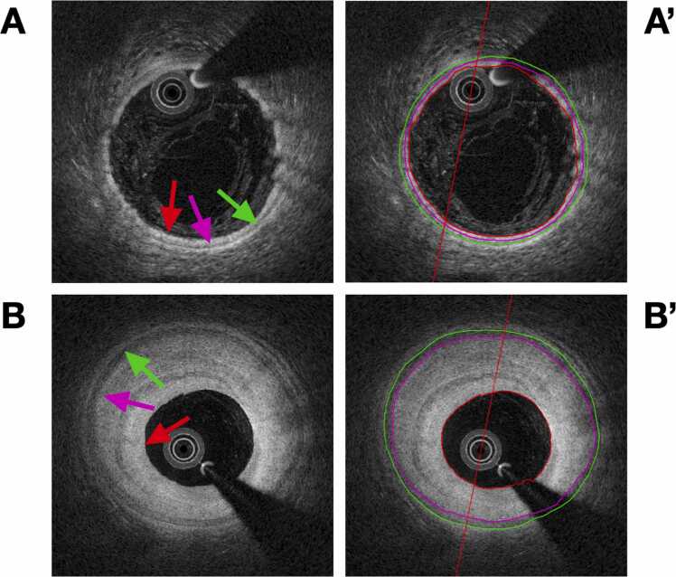
Background: Cardiac allograft vasculopathy (CAV) is a prevailing complication following heart transplantation. We aimed to investigate if CAV causes equal vascular remodeling in the major coronary arteries using quantitative optical coherence tomography (OCT) and to explore the prognostic potential of OCT-derived measurements from each coronary artery.
Methods: Sixty-four heart transplanted patients had a combined total of 114 full 3-vessel OCTs and coronary angiographies performed between 2013 and 2019. OCT pullbacks were categorized by angiographic CAV classification. Registration of disease progression was censored on July 1, 2022.
Results: OCT recordings were classified as follows: no significant CAV, n = 73; mild CAV, n = 18; moderate CAV, n = 13; and severe CAV, n = 10. From intercoronary comparison of severe CAV, we found significant differences by both average lumen/intima ratio (p < 0.0001) and average intima/media ratio (p < 0.0001). The left descending artery (LAD) showed increasingly smaller luminal areas and larger intimal areas within CAV groups compared with the remaining coronary arteries. No differences were seen between major coronary arteries without significant CAV. LAD derived average intima/media ratio (hazard ratio (HR): 3.39; 95% confidence interval (CI): 1.33-8.63; p = 0.01) and average lumen/intima ratio (HR: 2.77; 95% CI: 1.09-7.05; p = 0.03) were the strongest predictors of CAV progression.LAD predictions were superior to predictions based on all 3 coronary arteries.
Conclusions: LAD-derived OCT measurements were increasingly affected by CAV compared with the circumflex and right coronary artery. Average lumen/intima and intima/media ratios were the strongest predictors of CAV progression.
Keywords: cardiac allograft vasculopathy; coronary artery; heart transplantation; intravascular imaging; optical coherence tomography.
© 2024 International Society for Heart and Lung Transplantation.
Figures


References
-
- Clemmensen T.S., Munk K., Tram E.M., et al. Twenty years' experience at the Heart Transplant Center, Aarhus University Hospital, Skejby, Denmark. Scand Cardiovasc J. 2013;47:322–328. - PubMed
-
- Lund L.H., Khush K.K., Cherikh W.S., et al. The Registry of the International Society for Heart and Lung Transplantation: thirty-fourth adult heart transplantation report-2017; focus theme: allograft ischemic time. J Heart Lung Transplant. 2017;36:1037–1046. - PubMed
-
- Khush K.K., Cherikh W.S., Chambers D.C., et al. The International Thoracic Organ Transplant Registry of the International Society for Heart and Lung Transplantation: thirty-sixth adult heart transplantation report - 2019; focus theme: donor and recipient size match. J Heart Lung Transplant. 2019;38:1056–1066. - PMC - PubMed
-
- Tuzcu E.M., Kapadia S.R., Sachar R., et al. Intravascular ultrasound evidence of angiographically silent progression in coronary atherosclerosis predicts long-term morbidity and mortality after cardiac transplantation. J Am Coll Cardiol. 2005;45:1538–1542. - PubMed
-
- Torres H.J., Merello L., Ramos S.A., et al. Prevalence of cardiac allograft vasculopathy assessed with coronary angiography versus coronary vascular ultrasound and virtual histology. Transplant Proc. 2011;43:2318–2321. - PubMed
LinkOut - more resources
Full Text Sources
