Review
. 2025 Mar;48(2):e70021.
doi: 10.1002/jimd.70021.

Current Understanding of Pathogenic Mechanisms and Disease Models of Citrin Deficiency

Affiliations
Review

Current Understanding of Pathogenic Mechanisms and Disease Models of Citrin Deficiency

Denis Lacabanne et al. J Inherit Metab Dis. 2025 Mar.

Abstract

Citrin deficiency (CD) is a complex mitochondrial disease with three different age-related stages: neonatal intrahepatic cholestasis caused by CD (NICCD), failure to thrive and dyslipidemia caused by CD (FTTDCD), and type II citrullinemia (CTLN2), recently renamed adolescent and adult CD (AACD). While highly prevalent in the Asian population, CD is pan-ethnic and remains severely underdiagnosed. The disease is caused by the dysfunction or absence of the mitochondrial aspartate/glutamate carrier 2 (AGC2/SLC25A13), also known as citrin. Citrin deficiency results in a direct impairment of the malate-aspartate shuttle and the urea cycle, with expected knock-on effects on a multitude of other metabolic pathways, leading to a complicated pathophysiology. Here, we discuss our current knowledge of the molecular mechanism of substrate transport by citrin, including recent advances suggesting against its calcium regulation. We also discuss the different types of pathogenic variants found in CD patients and new insights into their pathogenic mechanisms. Additionally, we provide a summary and assessment of the efforts to develop preclinical models as well as treatments for the disease.

Keywords: citrin deficiency; disease models; mitochondrial transport; urea cycle disorders.


Conflict of interest statement

The authors declare no conflicts of interest.

Figures

FIGURE 1
FIGURE 1
Metabolic pathways related to citrin function. Schematic representation of the malate–aspartate shuttle (black), glycolysis (red), tricarboxylic acid (TCA) cycle (black), gluconeogenesis (blue), ammonia fixation/urea cycle (purple) and fatty acid oxidation (red). Citrin is shown in yellow together with the oxoglutarate carrier (OGC), ornithine carrier (ORC) and the carnitine/acylcarnitine carrier (CAC). The mitochondrial pyruvate carrier heterodimer (MPC) is shown in red/orange. The respiratory chain complexes 1 to 4 (CI–CIV), acyl‐CoA dehydrogenases (FI), electron transfer flavoprotein (FII), and ETF‐ubiquinone oxidoreductase (FIII) are shown in green and the dimer of ATP synthase in blue. PEP: phosphoenolpyruvate, NAD: nicotinamide adenine dinucleotide. NAD+ and NADH are highlighted in red.
FIGURE 2
FIGURE 2
Structural model of citrin. (A) Domain structure of citrin. In the N‐terminal domain, the EF‐hand‐containing mobile unit and static unit are shown in cyan and green, respectively. The calcium bound in EF‐hand 2 is shown as a green sphere. In the carrier domain, the three core elements are shown in blue, yellow and red and the three gate elements in gray. The C‐terminal domain is shown in orange. (B, C) Lateral and cytoplasmic view of the citrin structural model in the same color scheme as in (A).
FIGURE 3
FIGURE 3
The transport activity of reconstituted citrin is not regulated by calcium. (A) The calcium‐binding site of citrin in the presence of calcium (green sphere). (B) The mutated calcium binding site in the D66A/T68A/D70A/E77A mutant. (C) Purification of wild‐type citrin and the D66A/T68A/D70A/E77A mutant for in vitro assays. (D) Initial rates of substrate transport for wild‐type citrin and D66A/T68A/D70A/E77A, in the presence or absence of calcium. (E) Thermostability analysis for wild‐type citrin and D66A/T68A/D70A/E77A. (F) Oligomeric state of wild‐type citrin and D66A/T68A/D70A/E77A via size exclusion chromatography. (G) Super‐resolution mitochondrial imaging of a HAP1 cell line, where both endogenous citrin and aralar are knocked‐out, expressing transiently wild‐type citrin or D66A/T68A/D70A/E77A. (H) Line‐scan analysis of the relative fluorescence intensity of TOMM20, citrin or D66A/T68A/D70A/E77A and TIMM23 staining for the selected area shown in (F). Figure adapted from Tavoulari et al. [69].
FIGURE 4
FIGURE 4
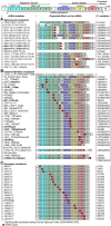
The wide range of pathogenic mutations of citrin. Positions of splicing, deletion, insertion, and nonsense variants reported in citrin deficiency, shown in the mRNA product (NM_014251.3). The red dots indicate the position of the substitutions. The color coding of the exons corresponds to the protein domains potentially translated, as shown in Figure 2. The potential translated protein outcome is indicated in the last column. The most recently reported variants (as in Table 1) are shown in bold. The effects of the splicing site variants were predicted using Human Splicing Finder [90].
FIGURE 5
FIGURE 5
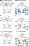
Effects of citrin missense variants on activity and mitochondrial localization. Positions of citrin missense variants in (A) the N‐terminal domain and (B) the carrier domain are highlighted in blue spheres. Novel variants most recently reported are shown in bold (also see Table 1). (C) Effect of citrin N‐terminal pathogenic variants on transport activity. (D) Effect of citrin carrier domain pathogenic variants on transport activity. For (C) and (D), residues with more than 50% activity are shown in green spheres, with 5%–50% activity with orange and with less than 5% activity in red. (E) Effect of citrin N‐terminal pathogenic variants on mitochondrial localization. (F) Effect of citrin carrier domain pathogenic variants on mitochondrial localization. In (E) and (F), mutants are hierarchically clustered for having primarily mitochondrial distribution (green spheres), mitochondrial distribution mixed with high levels of non‐mitochondrial (orange spheres) or primarily non‐mitochondrial distribution (red spheres).
FIGURE 6
FIGURE 6
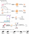
Generation procedures of cellular and mouse models of citrin deficiency. With the exception of patient‐derived induced Pluripotent Stem Cells (iPSCs), model design efforts have focused on knocking out the citrin gene. Ctrn, citrin; FACS, fluorescence‐activated cell sorting; KO, knock out; mGPD, mitochondrial glycerol‐3‐phosphate dehydrogenase; PBMCs, peripheral blood mononuclear cells. Image created with BioRender.com.

References

    1. Kobayashi K., Sinasac D. S., Iijima M., et al., “The Gene Mutated in Adult‐Onset Type II Citrullinaemia Encodes a Putative Mitochondrial Carrier Protein,” Nature Genetics 22 (1999): 159–163, 10.1038/9667. - DOI - PubMed
    1. Palmieri L., Pardo B., Lasorsa F. M., et al., “Citrin and Aralar1 Are Ca2+−Stimulated Aspartate/Glutamate Transporters in Mitochondria,” EMBO Journal 20 (2001): 5060, 10.1093/EMBOJ/20.18.5060. - DOI - PMC - PubMed
    1. Azzi A., Chappell J. B., and Robinson B. H., “Penetration of the Mitochondrial Membrane by Glutamate and Aspartate,” Biochemical and Biophysical Research Communications 29 (1967): 148–152, 10.1016/0006-291X(67)90556-6. - DOI - PubMed
    1. Dierks T. and Krämer R., “Asymmetric Orientation of the Reconstituted Aspartate/Glutamate Carrier From Mitochondria,” Biochimica et Biophysica Acta (BBA) ‐ Biomembranes 937 (1988): 112–126, 10.1016/0005-2736(88)90233-7. - DOI - PubMed
    1. Dierks T., Riemer E., and Krämer R., “Reaction Mechanism of the Reconstituted Aspartate/Glutamate Carrier From Bovine Heart Mitochondria,” Biochimica et Biophysica Acta (BBA) ‐ Biomembranes 943 (1988): 231–244, 10.1016/0005-2736(88)90555-X. - DOI - PubMed

MeSH terms

Supplementary concepts

LinkOut - more resources