. 2025 Jul;12(26):e2502073.
doi: 10.1002/advs.202502073. Epub 2025 Mar 27.

Elasticity-Driven Nanomechanical Interaction to Improve the Targeting Ability of Lipid Nanoparticles in the Malignant Tumor Microenvironment

Affiliations

Elasticity-Driven Nanomechanical Interaction to Improve the Targeting Ability of Lipid Nanoparticles in the Malignant Tumor Microenvironment

Eunhee Lee et al. Adv Sci (Weinh). 2025 Jul.

Abstract

The mechanical elasticity of lipid nanoparticles (LNPs) is crucial to their pharmaceutical performance. This study investigates how the mechanical interactions between LNPs, target cells, and macrophages affect the internalization of LNPs into target cells at tumor sites. According to our bio-mechanical study, drug-resistant breast cancer cells are stiffer than sensitive ones, while invasive cells are softer; similarly, protumoral M2 macrophages are softer than M1 macrophages. Softer LNPs show increased cellular uptake in breast cancer cells and macrophages, with enhanced engulfment in invasive cells and M2 macrophages. Additionally, the presence of M2 macrophages promotes greater LNP internalization by cancer cells, facilitating the malignant and invasive nature of cancer cells. In addition, because breast cancer cells engulf LNPs via an energy-efficient fusion pathway but LNPs in macrophages undergo clathrin-mediated endocytosis, LNPs are internalized more into cancer cells but not into M2. In orthotopic tumor models, softer LNPs penetrate tumors quickly, enhancing suppression, whereas stiffer LNPs permeate slowly but show prolonged retention in stiffer tumors, supporting antitumor efficacy with repeated dosing. These findings underscore the importance of mechanical interactions between LNPs, target cells, and macrophages in optimizing LNP delivery systems, offering insights for more effective designs.

Keywords: atomic force microscopy; cellular uptake; lipid nanoparticles; mechanical property; permeation.


Conflict of interest statement

The authors declare no conflict of interest.

Figures

Figure 1
Figure 1
Topographical and mechanical properties of the lipid nanoparticles (LNPs) obtained using atomic force microscopy (AFM). A) Topographical images acquired using AFM tapping mode with line profiles along the red lines. B) Uncompressed and compressed topographies of lipid nanoparticles (LNPs) derived from a 2D force–distance (fd) map with line profiles representing the cross‐sections. Each image is 700 × 700 nm with a resolution of 21.875 nm per direction. C) Representative 2D maps of the elastic moduli of LNPs under different stress regimes obtained from a 2D fd map. D) Average elastic moduli were measured at the centers and edges of DOPC+PBS and DOPC+Ca LNPs under various stress regimes (n = 5 per group). Data are presented as mean ± standard deviation (SD). Statistical significance was determined using a two‐tailed Student's t‐test; * p < 0.05, *** p < 0.005; ns: non‐significant (p > 0.05).
Figure 2
Figure 2
Elasticity of breast cancer cells and macrophages determined from the atomic force microscopy (AFM) indentation experiments. A–F) Representative force–indentation (ƒ–δ) curves from breast cancer cells (A–C) and macrophages (D–F). G,H) Average elasticity of the cell cortex (G) and body (H) for MDA‐MB‐231, MCF‐7, MCF‐7/ADR, RAW264.7, M1, and M2 cells measured at a trigger force of 5 nN for MDA‐MB‐231, MCF‐7, MCF‐7/ADR, RAW264.7, M1, and M2, respectively (n = 40 for each cell line). Error bars show standard error of the mean (SEM). Statistical significance was determined using a two‐tailed Student's t‐test; * p < 0.05, *** p < 0.005; &&& p < 0.005 versus RAW264.7; # p < 0.05, ### p < 0.005 versus M1; $$$ p < 0.005 versus M2.
Figure 3
Figure 3
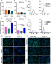
Differences in cellular uptake between low‐elasticity (DOPC+PBS) and high‐elasticity lipid nanoparticles (LNPs) (DOPC+Ca). A,B,D,E) Mean fluorescence intensity (MFI) obtained via flow cytometry representing the internalization rates of LNPs into breast cancer cells (A,B) and macrophages (D,E; n = 3 per group). Statistical significance was determined using a two‐tailed Student's t‐test. C,F) Representative graphs showing the number of breast cancer cells (C) and macrophages (F) versus fluorescence intensity. G,H) Fluorescence images of LNPs internalized into breast cancer cells (G) and macrophages (H; blue: DAPI, green: LNPs); * p < 0.05, ** p < 0.01, *** p < 0.005. Error bars show standard error of the mean (SEM).
Figure 4
Figure 4
Effect of interaction between breast cancer cells and macrophages on lipid nanoparticle (LNP) internalization. A) Schematic illustration of the measurement of LNP internalization in a coculture system of breast cancer cells and macrophages. Macrophages labeled with Cell Tracker Red CMTPX dye were visually discernible from breast cancer cells; thus, the amount of OG488‐DHPE‐labeled LNPs internalized into each cell type was measured using flow cytometry (FACS). B–I) Representative flow cytometry dot plots showing selective uptake of soft (B–E) and stiff (F–I) LNPs by different cell types in the coculture system. Colors represent MDA‐MB‐231, MCF‐7/ADR, M1, and M2 cells. J) Cumulative percentiles of fluorescence intensity generated by soft and stiff LNPs internalized into each cell type in each coculture system. Error bars show standard error of the mean (SEM). K) Relative uptake of soft and stiff LNPs into breast cancer cells compared with the total number of cells internalized with LNPs in each coculture system. Error bars represent standard error of the mean (SEM) (n = 5 per group). Statistical significance was determined using a two‐tailed Student's t‐test; * p < 0.05. L) Representative fluorescence images showing soft and stiff LNPs (green) internalized by breast cancer cells (blue) and macrophages (red).
Figure 5
Figure 5
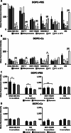
Endocytosis pathways of soft and stiff lipid nanoparticles (LNPs) in breast cancer cells and macrophages. A,B) Changes in the cellular uptake of soft (A) and stiff (B) LNPs upon treatment with chlorpromazine (clathrin‐mediated endocytosis inhibitor), dynasore (clathrin/caveolae‐mediated endocytosis inhibitor) and 4 °C (fusion inhibition; n = 3 per group). C,D) Identification of endocytosis pathways of soft (C) and stiff (D) LNPs in a coculture system when inhibiting endocytosis (n = 4 per group). Statistical significance was determined using a two‐tailed Student's t‐test; * p < 0.05, ** p < 0.01, *** p < 0.005 versus Control; # p < 0.05, ## p < 0.01, ### p < 0.005 4 °C versus 37 °C after 4 °C; Error bars show standard error of the mean (SEM).
Figure 6
Figure 6
Permeation of lipid nanoparticles (LNPs) labeled with green fluorescence into breast cancer spheroids with different Matrigel concentrations. Confocal images with line profiles along the yellow lines for A) 2.5% and B) 1.25% of Matrigel. Average fluorescence intensity at different Matrigel concentrations – C) 2.5% and D) 1.25% (n = 3 per group). Statistical significance was determined using a two‐tailed Student's test. Data are presented as mean ± standard deviation (SD); * p < 0.05 and *** p < 0.005 for the indicated comparison; ### p < 0.005 versus spheroid with 1.25% Matrigel.
Figure 7
Figure 7
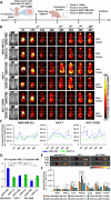
Biodistribution and antitumor efficacy of Cy5.5‐labeled DOPC+PBS.DOX and DOPC+Ca.DOX in orthotopic breast cancer models (n = 5). A) Schematic representation of the treatment regimen and tumor establishment in BALB/c nude mice. Ten intravenous injections were administered every 48 h after the tumor volume reached 100 mm3. B) In vivo fluorescence images showing the biodistribution of Cy5.5‐labeled DOPC+PBS and DOPC+Ca administered intravenously to mice with breast cancer. C) Fluorescence intensities of soft and stiff lipid nanoparticles (LNPs) observed in mice 2 h after each administration. D) Quantification of LNP accumulation by calculating the ratio of fluorescence intensities at 48 h after the 10th injection relative to the 1st injection. E) Fluorescence images of LNP‐treated tumors and major organs (brain, heart, liver, spleen, lung, and kidney) harvested from sacrificed mice at the experimental endpoint. F) Average radiant efficiencies from tumors and major organs harvested from LNP‐treated mice. Statistical significance for C,D,F) was determined using a two‐tailed Student's t‐test; * p < 0.05, ** p < 0.01, *** p < 0.005. Error bars represent standard deviation (SD).
Figure 8
Figure 8
Antitumor efficacy of DOPC+PBS.DOX and DOPC+Ca.DOX in orthotopic breast cancer models (n = 5). A) Relative tumor volume compared with the initial tumor volume in each treatment group over time; * p < 0.05, *** p < 0.005 versus saline; # p < 0.05, ### p < 0.005 versus free DOX; $ p < 0.05 versus DOPC+PBS.DOX. B) Photograph of resected tumors from breast tumor‐bearing mice treated with saline, free DOX, DOPC+PBS.DOX, or DOPC+Ca.DOX. C) Tumor size measured in tumor‐bearing mice before the treatment and after resection at the experimental endpoint; * p < 0.05, *** p < 0.005. Statistical significance for A,C) was determined using a two‐tailed Student's t‐test. Error bars represent standard deviation (SD).
Figure 9
Figure 9
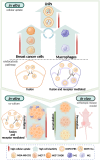
Schematic illustration depicting the cellular uptake and internalization mechanisms influenced by the mechanical properties of lipid nanoparticles (LNPs), breast cancer cells, and macrophages.

Similar articles

Cited by

References

    1. a) Åberg C., Piattelli V., Montizaan D., Salvati A., Nanoscale 2021, 13, 17530; - PMC - PubMed
    2. b) Xia Q., Huang J., Feng Q., Chen X., Liu X., Li X., Zhang T., Xiao S., Li H., Zhong Z., Xiao K., Int. J. Nanomed. 2019, 14, 6957. - PMC - PubMed
    1. Kiio T. M., Park S., J. Pharm. Invest. 2021, 51, 35.
    1. Zou D., Wu Z., Yi X., Hui Y., Yang G., Liu Y., Wang H., Brooks A., Wang H., Liu X., Xu Z. P., Roberts M. S., Gao H., Zhao C.‐X., Proc. Natl. Acad. Sci. U. S. A. 2023, 120, 2214757120. - PMC - PubMed
    1. a) Ma X., Yang X., Li M., Cui J., Zhang P., Yu Q., Hao J., Langmuir 2021, 37, 11688; - PubMed
    2. b) Hui Y., Yi X., Wibowo D., Yang G., Middelberg A. P. J., Gao H., Zhao C. X., Sci. Adv. 2020, 6, aaz4316. - PMC - PubMed
    1. Hui Y., Yi X., Hou F., Wibowo D., Zhang F., Zhao D., Gao H., Zhao C. X., ACS Nano 2019, 13, 7410. - PubMed

LinkOut - more resources