

The Effect of Partial Sleep Deprivation on Retinal Microvasculature in Myopia With Different Axial Lengths
- PMID: 40146130
- PMCID: PMC11954537
- DOI: 10.1167/iovs.66.3.57
The Effect of Partial Sleep Deprivation on Retinal Microvasculature in Myopia With Different Axial Lengths
Abstract
Purpose: To investigate the effects of partial sleep deprivation (PSD) on the retinal microvasculature in individuals with myopia, using optical coherence tomography angiography (OCTA).
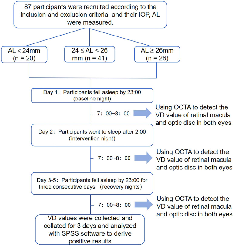
Methods: In total, 87 right eyes were categorized by axial length (AL) into three groups: A (AL < 24 mm, n = 20), B (24 ≤ AL < 26 mm, n = 41), and C (AL ≥ 26 mm, n = 26). Participants underwent macular (6 mm × 6 mm) and optic disc (4.5 mm × 4.5 mm) OCTA scans. Vessel density (VD) parameters-including superficial capillary plexus VD (SCP-VD), deep capillary plexus VD (DCP-VD), radial peripapillary capillary VD (RPC-VD), inside disc RPC VD (iVD), and peripapillary RPC VD (ppVD)-were compared at baseline, before and after PSD, and following 3 days of regular sleep.
Results: Compared with group A, groups B and C had lower baseline DCP-VD in all subregions (P < 0.05), except for the perifovea-inferior area, where only groups C and A showed significant differences (P = 0.001). After PSD, group C showed a decrease in SCP-VD in certain subregions of the parafovea (P = 0.013 and P = 0.022 for parafovea-temporal and parafovea-inferior, respectively), along with an increase in ppVD (P = 0.012). All VD parameters recovered after 3 days of regular sleep (P < 0.05).
Conclusions: The retinal microvasculature of myopic eyes with an AL of ≥26 mm show greater susceptibility to PSD than those with an AL of <26 mm. However, short-term PSD effects can be restored by setting a regular sleep schedule.
Conflict of interest statement
Disclosure:
Figures


Similar articles
-
The Effect of Axial Length on Macular Vascular Density in Eyes with High Myopia.Rom J Ophthalmol. 2025 Jan-Mar;69(1):88-100. doi: 10.22336/rjo.2025.15. Rom J Ophthalmol. 2025. PMID: 40330963 Free PMC article.
-
The vascular densities of the macula and optic disc in normal eyes from children by optical coherence tomography angiography.Graefes Arch Clin Exp Ophthalmol. 2020 Feb;258(2):437-444. doi: 10.1007/s00417-019-04466-0. Epub 2019 Nov 15. Graefes Arch Clin Exp Ophthalmol. 2020. PMID: 31732811
-
Longitudinal changes in optical coherence tomography angiography characteristics in normal-tension glaucoma with or without high myopia.Acta Ophthalmol. 2024 Aug;102(5):e762-e773. doi: 10.1111/aos.16644. Epub 2024 Jan 26. Acta Ophthalmol. 2024. PMID: 38279584
-
Analysis of Retinal Microvasculature Features in Amblyopic Eyes: A Meta-Analysis.Ophthalmic Res. 2023;66(1):131-143. doi: 10.1159/000526531. Epub 2022 Aug 23. Ophthalmic Res. 2023. PMID: 35998587
-
Evaluation of Posterior Ocular Blood Flow in Diabetic Retinopathy Patients Without Macular Edema Using Optical Coherence Tomography Angiography.Photodiagnosis Photodyn Ther. 2023 Dec;44:103777. doi: 10.1016/j.pdpdt.2023.103777. Epub 2023 Sep 3. Photodiagnosis Photodyn Ther. 2023. PMID: 37669724 Review.
References
-
- Dolgin E. The myopia boom. Nature. 2015; 519(7543): 276–278. - PubMed
-
- Morgan IG, Ohno-Matsui K, Saw SM. Myopia. Lancet. 2012; 379(9827): 1739–1748. - PubMed
-
- Morgan IG, French AN, Ashby RS, et al. .. The epidemics of myopia: aetiology and prevention. Prog Retin Eye Res. 2018; 62: 134–149. - PubMed
-
- Yaprak AC, Yaprak L.. Retinal microvasculature and optic disc alterations in non-pathological high myopia with optical coherence tomography angiography. Graefes Arch Clin Exp Ophthalmol. 2021; 259(11): 3221–3227. - PubMed
MeSH terms
LinkOut - more resources
Full Text Sources
Miscellaneous
