Review
. 2025 Mar 28;136(7):752-772.
doi: 10.1161/CIRCRESAHA.124.325685. Epub 2025 Mar 27.

Biophysical and Biochemical Roles of Shear Stress on Endothelium: A Revisit and New Insights

Affiliations
Review

Biophysical and Biochemical Roles of Shear Stress on Endothelium: A Revisit and New Insights

Chak Kwong Cheng et al. Circ Res. .

Abstract

Hemodynamic shear stress, the frictional force exerted by blood flow on the endothelium, mediates vascular homeostasis. This review examines the biophysical nature and biochemical effects of shear stress on endothelial cells, with a particular focus on its impact on cardiovascular pathophysiology. Atherosclerosis develops preferentially at arterial branches and curvatures, where disturbed flow patterns are most prevalent. The review also highlights the range of shear stress across diverse human arteries and its temporal variations, including aging-related alterations. This review presents a summary of the critical mechanosensors and flow-sensitive effectors that respond to shear stress, along with the downstream cellular events that they regulate. The review evaluates experimental models for studying shear stress in vitro and in vivo, as well as their potential limitations. The review discusses strategies targeting shear stress, including pharmacological approaches, physiological means, surgical interventions, and gene therapies. Furthermore, the review addresses emerging perspectives in hemodynamic research, including single-cell sequencing, spatial omics, metabolomics, and multiomics technologies. By integrating the biophysical and biochemical aspects of shear stress, this review offers insights into the complex interplay between hemodynamics and endothelial homeostasis at the preclinical and clinical levels.

Keywords: atherosclerosis; endothelial cells; exercise; hemodynamics; multiomics.


Conflict of interest statement

None.

Figures
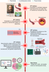
Figure 1.
Figure 1.
A brief history of hemodynamic study. A review of the past, contemporary, and future contexts of hemodynamic studies on endothelial cells (ECs). Leonardo Da Vinci’s portrait was created by Leonardo AI. Processing software: Biorender.
Figure 2.
Figure 2.
Biophysical features of hemodynamic flow and shear stress. A, Exertion of various forms of mechanical stress on endothelial cells (ECs) by hemodynamic flow, including shear stress, blood pressure, and tensile strain. B, Types. C, Dynamics. D, Directions of hemodynamic flow experienced by ECs. E, The relationship between hemodynamic parameters (ie, shear rate, shear stress, and viscosity), and corresponding atherosclerosis risk. F, Variation of shear stress in the upstream, midstream, and downstream portions of atherosclerotic plaque. G, Shear stress ranges and prevalence of atherosclerosis in different types of human arteries. H, Variation of shear stress magnitudes between species, such as the human and mouse. I, A decline in shear stress intensities has been observed in the common carotid arteries of both male and female individuals during the aging process. Processing software: Biorender.
Figure 3.
Figure 3.
Biochemical effects of hemodynamic shear stress. Mechanosensors and mechanosensitive structures on the apical and basal surfaces and at cell-cell junctions of endothelial cells (ECs) convert biomechanical stimuli of shear stress to biochemical signals, which modulate endothelial and vascular events through various pathways and the action of different effectors (eg, proteins, microRNAs [miRNAs], and long noncoding RNAs [lncRNAs]). Steady laminar shear stress (LSS) and oscillatory shear stress (OSS) have been shown to activate different pathways, thereby eliciting endothelial and vascular effects. Processing software: Biorender. AKT indicates protein kinase B; AMPK, adenosine monophosphate-activated protein kinase; BMP, bone morphogenetic protein; EndoMT, endothelial‐to‐mesenchymal transition; eNOS, endothelial NO synthase; ERK, extracellular signal-regulated kinase; FAK, focal adhesion kinase; GPCR, G-protein–coupled receptor; HIF1α, hypoxia-inducible factor 1α; KLF, Krüppel-like factor; lncRNA, long noncoding RNA; MEK5, mitogen-activated protein kinase kinase 5; miRNA, microRNA; NADPH, nicotinamide adenine dinucleotide phosphate; NF-κB, nuclear factor-κB; NOTCH1, neurogenic locus notch homolog protein 1; NRF2, nuclear factor erythroid 2–related factor 2; P2X4, P2X purinoreceptor 4; PECAM1, platelet endothelial cell adhesion molecule-1; PI3K, phosphoinositide 3-kinase; PIEZO1, piezo type mechanosensitive ion channel component 1; ROS,reactive oxygen soecies; SIRT1, sirtuin 1; SMAD1/5, mothers against decapentaplegic homolog 1/5; SOX, SRY-box transcription factor; TAZ, transcriptional coactivator with PDZ-binding motif; UCP2, uncoupling protein 2; VE, vascular endothelial; VEGFR2/3, vascular endothelial growth factor receptor 2/3; and YAP, yes-associated protein.
Figure 4.
Figure 4.
In vitro and in vivo experimental models for hemodynamic study. A, A parallel-plate flow chamber connected to an ibidi pump system is used to expose endothelial cells (ECs) to differential flow generated by a computer. B, Cone-and-plate viscometer, where ECs are exposed to differential flow generated by a rotating Teflon cone. C, Orbital shaker, where ECs are concurrently exposed to antiatherogenic and proatherogenic flows. D, Microfluidic devices developed for use in a range of hemodynamic studies. E, Flow patterns and shear stress magnitudes at different sites of the aorta. F, Mouse models commonly used for hemodynamic studies. G, Partial carotid ligation model is used to induce disturbed flow in the left carotid artery (LCA) by surgically ligating the external carotid artery (ECA), internal carotid artery (ICA), and occipital artery (OA), while the right carotid artery (RCA) serves as a control. H, Carotid cuff implantation model involves the implantation of a constrictive cuff to induce low steady laminar shear stress (LSS), increasing LSS, and low oscillatory shear stress (OSS) along the RCA, while the LCA serves as a control. Processing software: Biorender. AAV9 indicates adeno-associated virus 9; LSA, left subclavian artery; PCSK9, proprotein convertase subtilisin/kexin type 9; RSA, right subclavian artery; and STA, superior thyroid artery.
Figure 5.
Figure 5.
Strategies targeting hemodynamic shear stress. There are 4 main ways in which pharmacological, genetic, physiological, and surgical strategies can target hemodynamic shear stress. These are (1) increasing steady laminar shear stress (LSS) or mimicking its beneficial effects, (2) diminishing oscillatory shear stress (OSS) or its harmful effects, (3) targeting downstream effectors, and (4) modulating downstream events induced by shear stress. The rational design of combination therapy regimens has the potential to optimize hemodynamic performance. Processing software: Biorender.
Figure 6.
Figure 6.
New perspectives for hemodynamic studies. A, Proposed major endothelial cell (EC) subpopulations and their corresponding phenotypes based on single-cell sequencing results from different hemodynamic studies. B, Integration of existing bulk and single-cell omics data as a future perspective for hemodynamic research. C, Future perspectives for hemodynamic studies, with a particular focus on single-cell and spatial omics. Processing software: Biorender. 3D indicates 3-dimensional; EndoMT, endothelial‐to‐mesenchymal transition; and miRNA, microRNA.

References

    1. Tamargo IA, Baek KI, Kim Y, Park C, Jo H. Flow-induced reprogramming of endothelial cells in atherosclerosis. Nat Rev Cardiol. 2023;20:738–753. doi: 10.1038/s41569-023-00883-1 - PMC - PubMed
    1. Sterpetti AV. Cardiovascular research by Leonardo da Vinci (1452–1519). Circ Res. 2019;124:189–191. doi: 10.1161/CIRCRESAHA.118.314253 - PubMed
    1. Caro CG. Discovery of the role of wall shear in atherosclerosis. Arterioscler Thromb Vasc Biol. 2009;29:158–161. doi: 10.1161/ATVBAHA.108.16673 - PubMed
    1. Cheng CK, Huang Y. Vascular endothelium: the interface for multiplex signal transduction. J Mol Cell Cardiol. 2024;195:97–102. doi: 10.1016/j.yjmcc.2024.08.004 - PubMed
    1. Libby P, Buring JE, Badimon L, Hansson GK, Deanfield J, Bittencourt MS, Tokgözoğlu L, Lewis EF. Atherosclerosis. Nat Rev Dis Primers. 2019;5:56. doi: 10.1038/s41572-019-0106-z - PubMed