. 2025 Mar 27;16(1):3003.
doi: 10.1038/s41467-025-58190-x.

Proapoptotic Bcl-2 inhibitor as potential host directed therapy for pulmonary tuberculosis

Affiliations

Proapoptotic Bcl-2 inhibitor as potential host directed therapy for pulmonary tuberculosis

Medha Singh et al. Nat Commun. .

Abstract

Mycobacterium tuberculosis establishes within host cells by inducing anti-apoptotic Bcl-2 family proteins, triggering necrosis, inflammation, and fibrosis. Here, we demonstrate that navitoclax, an orally bioavailable, small-molecule Bcl-2 inhibitor, significantly improves pulmonary tuberculosis (TB) treatments as a host-directed therapy. Addition of navitoclax to standard TB treatments at human equipotent dosing in mouse models of TB, inhibits Bcl-2 expression, leading to improved bacterial clearance, reduced tissue necrosis, fibrosis and decreased extrapulmonary bacterial dissemination. Using immunohistochemistry and flow cytometry, we show that navitoclax induces apoptosis in several immune cells, including CD68+ and CD11b+ cells. Finally, positron emission tomography (PET) in live animals using clinically translatable biomarkers for apoptosis (18F-ICMT-11) and fibrosis (18F-FAPI-74), demonstrates that navitoclax significantly increases apoptosis and reduces fibrosis in pulmonary tissues, which are confirmed in postmortem analysis. Our studies suggest that proapoptotic drugs such as navitoclax can potentially improve pulmonary TB treatments, reduce lung damage / fibrosis and may be protective against post-TB lung disease.


Conflict of interest statement

Competing interests: The authors declare no competing interests.

Figures

Fig. 1
Fig. 1. Navitoclax treatment in mouse model of pulmonary tuberculosis at human equipotent dosing.
M. tuberculosis-infected mice were randomly allocated to receive standard TB treatment (R, rifampin; H, isoniazid; Z, pyrazinamide) with or without navitoclax at human equipotent dosing via oral gavage. a TB treatments were initiated three weeks after a high dose aerosol infection. Bacterial burden [colony-forming unit (CFU) per mL (log10) from whole lung] after three weeks of treatment (n = 3 untreated mice, 3 navitoclax (alone) treated mice, 4 RHZ treated mice and 4 RHZ + navitoclax treated mice). When combined with the standard TB treatment (RHZ + navitoclax), there was a significant (albeit modest) reduction in the bacterial burden compared to the standard treatment alone (RHZ) (P < 0.01). b Hematoxylin & Eosin (H&E) stained lung sections of mice demonstrating lung pathology. c The bar graph represents the percentage of affected lung tissue regions from H&E-stained lung tissue sections (n = 3 tissue sections from untreated group and 5 tissue sections from RHZ and RHZ + Navitoclax treated group from 2 animals per group). The addition of navitoclax also improved lung pathology, with a significant decrease in the percentage of lesion affected lung regions (P = 0.02). d TB treatments were initiated six weeks after a low dose aerosol infection with M. tuberculosis, when fibrosis is well established (chronic model). Bacterial burden [colony-forming unit (CFU) per mL (log10) from whole lung] after eight weeks of treatment (n = 5 mice/group) are shown. Mice receiving navitoclax in addition to the standard TB treatment at eight-weeks of treatment had significantly lower bacterial burden (1.2 log10 reduction) (P = 0.04). e Lung tissue H&E sections were used for quantification of necrosis using Trainable Weka Segmentation 3D plugin available in ImageJ FIJI (n = 5 sections from each group). f Percent necrotic area was quantified for each lesion and shown as bar graph. Necrotic areas were substantially lower in mice treated with adjunctive navitoclax versus RHZ alone (P = 0.01). For CFU, data are represented as mean ± standard deviation and statistical comparisons were made using the student t-test. For affected and necrotic lung tissue regions, data are represented as median ± interquartile range. Statistical comparisons were made using the Mann-Whitney U one-tailed test or the student two-tailed t-test (CFU data).
Fig. 2
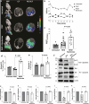
Fig. 2. Apoptosis quantification using imaging in live M. tuberculosis-infected mice and postmortem analysis.
TB treatments were initiated three weeks after a high dose aerosol infection. a Maximum intensity projection (MIP) and transverse 18F-ICMT-11 PET/CT from representative M. tuberculosis-infected mice from the different treatment arms, two weeks after initiation of TB treatments. Quantification of pulmonary 18F-ICMT-11 PET signal as percent injected dose/mL (%ID/mL) (panel b) and area under curve (AUC) (panel c) which was significantly higher in animals treated with the standard TB treatment in addition to navitoclax versus those receiving the standard treatment alone (P = 0.01; n = 3 untreated mice, 4 RHZ treated mice and 4 RHZ + navitoclax treated mice) are shown. d Flow cytometry of single-cell suspensions to analyse the percentage of Annexin V positive cells (n = 3 animals per group; samples were acquired in duplicates for some groups). e Lung tissue homogenate caspase 3 activity is shown (n = 4 animals per group). Apoptosis markers, Annexin V (P < 0.01) and caspase 3 (P = 0.01) were significantly higher in mice receiving navitoclax plus standard TB treatment versus standard TB treatment alone. Levels of anti-apoptotic proteins Bcl2 and Bcl-xl (panels fh), and pro-apoptotic proteins Bim, Bid, Cytochrome C (Cyt C) (panels f, ik) from lung tissue homogenates using GAPDH as an internal control are shown (n = 4 animals per group). Bcl-2 and Bcl-xl protein levels were significantly lower (P = 0.01 and P = 0.04, respectively), and levels of Bim, Bid and Cyt C were significantly higher (P = 0.01, P = 0.04, and P = 0.01, respectively) in animals receiving adjunctive navitoclax versus standard TB treatment alone. Data are represented as median ± interquartile range. Statistical comparisons were made using the Mann-Whitney U one-tailed test.
Fig. 3
Fig. 3. High-dimensional flow cytometry in lung tissues.
Isoniazid with or without navitoclax were initiated three weeks after a high dose aerosol infection. Cell suspensions from lung tissues of M. tuberculosis-infected mice from the different treatment arms after exclusion of debris and doublets were analysed, two weeks after initiation of TB treatments. ac Distribution of immune cells (CD45+) in the different treatment arms is shown. d Percentage of cells positive for intracellular expression of cleaved caspase 3 is shown. Five animals were used for each group. The addition of navitoclax led to a significant increase in apoptosis in several myeloid / macrophage lineage cells, two weeks after treatment initiation (P < 0.01). e TB treatments (RHZ with or without navitoclax) were initiated six weeks after a low dose aerosol infection with M. tuberculosis, when fibrosis is well established (chronic model). TGF-β levels (n = 4 mice/group) were assessed in CD45+ immune cell populations (alveolar macrophage and interstitial macrophage) and CD45- non-immune cell populations (epithelial, endothelial and fibroblast) using flow cytometry. TGF-β levels in the lung tissues of navitoclax + RHZ (versus RHZ alone) treated animals were significantly lower in immune and non-immune cells (P = 0.03), four weeks after treatment initiation. Data are represented as median ± interquartile range. Statistical comparisons were made using the Mann-Whitney one-tailed U test.
Fig. 4
Fig. 4. Immunohistochemistry in lung tissues.
Fixed lung tissues from M. tuberculosis-infected mice from the different treatment arms, three weeks after initiation of TB treatments (n = 4 sections/group; 2 sections per animal). TB treatments were initiated three weeks after a high dose aerosol infection. a H&E-stained images and immunostained panels - cleaved caspase 3, CD68 and merged are shown (40x magnification, scale bar 25 μm). b Pearson’s coefficient to quantify colocalization of cleaved caspase 3 and CD68 is shown. There is significantly higher colocalization of cleaved caspase 3 and CD68 in mice receiving navitoclax plus standard TB treatment versus those receiving standard treatment alone (P = 0.03). Cumulative mean fluorescence intensity (MFI) for (c), cleaved caspase 3 (RHZ versus RHZ + navitoclax, P = 0.01) and (d), CD68+. Data are represented as median ± interquartile range. Statistical comparisons were made using the Mann-Whitney U one-tailed test.
Fig. 5
Fig. 5. Fibrosis imaging in live M. tuberculosis-infected mice.
TB treatments were initiated six weeks after a low dose aerosol infection with M. tuberculosis, when fibrosis is well established (chronic model). a Maximum intensity projection (MIP) and transverse 18F-FAPI-74 PET/CT from representative M. tuberculosis-infected mice from the different treatment arms, two weeks after initiation of TB treatments. Quantification of pulmonary 18F-FAPI-74 PET signal as lesion to blood ratio (panel b) and heatmaps representing area under the curve (AUClesion/blood) ratio (panel c) (n = 4 mice/group) are shown. A significantly lower AUClesion/blood was observed in animals receiving adjunctive navitoclax versus standard TB treatment alone (P = 0.03). Data are represented as median ± interquartile range. Statistical comparisons were made using the Mann-Whitney U one-tailed test.
Fig. 6
Fig. 6. Postmortem studies to quantify lung tissue fibrosis.
Fixed lung tissues from M. tuberculosis-infected mice (chronic model) from the different treatment arms were used two and four weeks after initiation of TB treatments (n = 6 sections per group). Representative Masson’s trichrome stained sections (panel a) and quantification (panel b) is shown. c Soluble collagen was quantified in whole lung lysates (n = 4 animals/group). A significantly lower pulmonary fibrosis [Masson’s trichrome staining (P < 0.01) and soluble collagen levels (P = 0.02)] was observed in animals receiving adjunctive navitoclax versus standard TB treatment alone. Data are represented as median ± interquartile range. Statistical comparisons were made using the Mann-Whitney U one-tailed test.

Update of

References

    1. WHO. Global Tuberculosis Report (2024).
    1. Malik, Z. A., Iyer, S. S. & Kusner, D. J. Mycobacterium tuberculosis phagosomes exhibit altered calmodulin-dependent signal transduction: contribution to inhibition of phagosome-lysosome fusion and intracellular survival in human macrophages. J. Immunol.166, 3392–3401 (2001). - PubMed
    1. Pan, H. et al. Ipr1 gene mediates innate immunity to tuberculosis. Nature434, 767–772 (2005). - PMC - PubMed
    1. Aguiló, N., Marinova, D. & Martín, C. J. P. ESX-1-induced apoptosis is involved in cell-to-cell spread of Mycobacterium tuberculosis. Cell Microbiol. 15, 1994–2005 (2013). - PubMed
    1. Behar, S. M. et al. Apoptosis is an innate defense function of macrophages against. Mucosal Immunol.4, 279–287 (2011). - PMC - PubMed

MeSH terms

LinkOut - more resources