. 2025 Mar 28;15(1):10669.
doi: 10.1038/s41598-025-94064-4.

Factor H-related 2 levels dictate FHR dimer composition

Affiliations

Factor H-related 2 levels dictate FHR dimer composition

Bert R J Veuskens et al. Sci Rep. .

Abstract

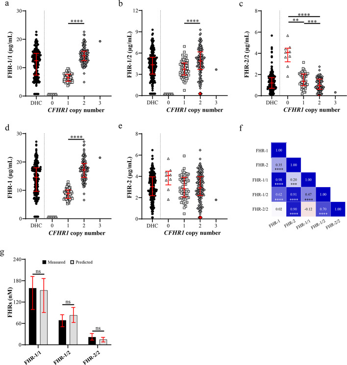
Factor H-related (FHR) protein 1 and 2 form dimers resulting in FHR-1 and -2 homodimers, and FHR-1/2 heterodimers. Dimerization is hypothesized to further increase their antagonistic function with complement regulator factor H (FH). So far, only FHR-1 homodimers and FHR-1/2 heterodimers could be quantified in a direct way. With the reported genetic associations between CFHR2 and complement-related diseases such as age related macular degeneration and C3-glomerulopathy, direct assessment of FHR-2/2 levels determining the dimer distribution of FHR-1 and -2 is needed to further elucidate their role within complement regulation. Therefore, novel in-house generated FHR-2 antibodies were used to develop a specific ELISA to enable direct quantification of FHR-2 homodimers. Allowing for the first time the accurate measurement of all FHR-1 and -2 containing dimers in a large cohort of healthy donors. By using native FHR-1 and -2 or deficient plasma, we determined the stability, kinetics and distribution of FHR-1 and -2 dimers. Additionally, we show how genetic variants influence dimer levels. Our results confirm a rapid, dynamic, dimer formation in plasma and show FHR-1/2 dimerization rearches a distribution equilibrium that is limited by the relative low levels of FHR-2 in relation to its dimerization partner FHR-1.

Keywords: Complement system; Dimerization; FHR-1; FHR-2.


Conflict of interest statement

Declarations. Competing interests: MCB, TWK and RBP are co-inventors of patents and patents applications describing potentiating anti-FH antibodies and uses thereof. All other authors declare no conflict of interest. Ethical approval: After consultation with the ethical board of Sanquin Research, Amsterdam, The Netherlands, a system was established for obtaining blood samples for scientific research (no approval number available). This volunteer system is organized according to Dutch regulations and according to the Declaration of Helsinki. This volunteer system certifies, among others, that: Blood samples used for scientific studies by researchers of the Sanquin Research department were drawn from healthy, anonymized volunteers with written informed consent; No personal characteristics of the volunteers are registered; The volunteers nor those taking the samples know for what project specific samples are used; Allowed annual sample volume and frequency of donation were established after consultation with Sanquin Medical Secretary. Standard operating procedures are available upon request.

Figures

Fig. 1
Fig. 1
Characterization of FHR-2 monoclonal antibodies (mAbs). (a) An overview illustrating the genetic similarity between FHR-1, -2, and FHR-5. (b) Cross-reactivity analysis of the αFHR-2 mAbs. Wells were coated with the indicated αFHR-2 mAb and incubated with biotinylated plasma-derived FH (pdFH) or recombinant human FHR (rhFHR) proteins (10 nM). Current non-specific αFHR-2 mAbs (αFHR-2.1, αFHR-2.4) and an irrelevant isotype mAb (neg. contrl.) were included as controls. Absorption levels above 0.1 (dotted line) were considered indicative of cross-reactivity. Some test conditions resulted in absorption levels exceeding the upper limit of quantification, set at 3.0. (ce) Western blot analysis of immunoprecipitation using αFHR-2.11 to αFHR-2.15 in normal human serum (c), CFHR3/CFHR1 deficient serum (determined via MLPA) (d), and serum deficient for FHR-2 (previously determined via gene sequencing) (e). All three immunoblots were stained using αFHR-2.1 (cross-reactive for FHR-1 and -2) to verify specificity for native FHR-2 and exclude cross-reactivity for native FHR-1. (f) Competition ELISA including selective αFHR-2 mAbs. Biotinylated rhFHR-2 (0.1 µg/mL) was pre-incubated with αFHR-2 mAbs (10 µg/mL) for twenty minutes before adding to the ELISA plates coated with indicated mAbs. Binding of biotinylated rhFHR-2 is expressed as relative binding of biotinylated rhFHR-2 in the presence of an isotype control. Bars represent the mean of two independent replicates with error bars indicating the SD. ELISAs and Western blots are representatives of three independent experiments.
Fig. 2
Fig. 2
ELISA validation and calibration. (a) Selection of monoclonal antibody (mAb) sandwich combination to enable detection of FHR-2/2 homodimers in normal human serum (NHS). Combinations of the same mAb (bolt bordered squares) are of interest based on the principal only a homodimer presents a single FHR-2 epitope twice. ELISA plates were coated with a FHR-2 specific αFHR-2 mAb and incubated with 20% (v/v) NHS. Next, bound FHR-2 was detected using indicated biotinylated αFHR-2 mAb. Absorption levels above 0.1 were considered indicative of antigen detection. Validation of FHR-2/2 (b) and FHR-1/2 (c) assay specificity using NHS and serum deficient for FHR-1 or -2. The FHR-2 specific αFHR-2.11 mAb was coated on ELISA plates and incubated with either NHS and serum deficient for FHR-1 or -2 (4%, (v/v)). Next, bound target protein was detected using biotinylated αFH.02 (αFH mAb cross-reactive with FHR-1) or αFHR-2.11 for FHR-1/2 and -2/2 dimers, respectively. (d) FHR-2/2 assay calibration of an NHS standard using rhFHR-2. For the calibration of the FHR-1/2 ELISA, seven mixes with different volumetric ratios (mix A: 95–5%, B: 85–15%, C: 75–25%, D: 50–50%, E: 25–75%, F: 15–85%, G: 5–95% (v/v)) of a plasma deficient in FHR-2 or -1 respectivaly were mixed and incubated at 37 °C for six hours to allow the formation of FHR-1/2 heterodimers. (e) NHS standard pool calibrated for FHR-1/2 using each mix as standard. FHR-1/2 levels in each mix were based on the average decline in FHR-1/1 and -2/2. Dotted line represents the mean of mix C-G with the grey area indicating the mean ± 15%. (f) Levels of all three FHR-1 and -2 dimer species in each mix (start concentration FHR-1/1: 273.84 nM; start concentration FHR-2/2: 81.20 nM) after incubation at 37 °C. (ad) n= 3, representative is shown. (bf) Symbols represent mean of multiple measurements with error bars indicating the SD.
Fig. 3
Fig. 3
Insights into FHR-1 and -2 dimerization. (a) FHR-1/2 ELISA was used to show the rapid formation of FHR-1/2 dimers over time when incubating equimolar amount of FHR-1 and FHR-2 at 37 °C. A plasma pool deficient in FHR-1 or FHR-2 was used as source for FHR-2/2 and FHR-1/1 respectively. FHR-1/2 levels were measured at 4 °C to halt further dimer formation. (b) Impact of pH on the stability of FHR-1 and -2 dimers. Normal human serum (NHS, 3% v/v) was incubated at the indicated pH using an isotonic 40 mM BisTrispropane buffer. pH 8.5 was used as reference, resembling the pH of the in-house dimer ELISAs. (c) Impact of sodium chloride on dimer stability. NHS (3%, (v/v) was incubated with increasing concentrations of sodium chloride ranging from 0.0625–2 M. 125 mM NaCl was used as a 100% reference. Symbols represent mean of three separate measurements with error bars indicating the SD.
Fig. 4
Fig. 4
Reference intervals of FHR-1, -2 and their dimers in healthy donors. Reference levels of (a) FHR-1/1, (b) FHR-1/2, (c) FHR-2/2 and total (calculated) levels of FHR-1 (d) and -2 (e) in serum of 201 Dutch healthy volunteers. Donors are categorized based on their copy number of CFHR1 as determined with MLPA. Total levels of FHR-1 and -2 were calculated using measured levels of FHR-1 and -2 dimers. Red colored diamonds are donors identified to lack FHR-2 as previously validated by van Beek et al.. (f) Pearson correlation of FHR-1 and -2 and their dimer species. (g) Comparison of measured and predicted FHR-1 and -2 dimers in 190 Dutch healthy controls. Predicted levels were determined applying the calculations for when reaching a distribution equilibrium,. The Shapiro–Wilk test was used to test for normal distribution of the population. (a, b, d) Unpaired t-test with Welch’s correction, (c) the Kruskal Wallis test, (e) the Brown-Forsythe and Welch ANOVA tests, and lastly (g) the Chi-squared test were used to test for significant differences. The Dunn’s test (c) and (d) the Dunnett’s T3 multiple comparisons test were used to correct for multiple testing. (a, b, c, d, e) Donors deficient for FHR-1 or lacking FHR-2 were excluded in the statistical analysis. Symbols represent the mean of two measurements with error bars indicating the median with interquartile range. (* p < 0.05; ** p < 0.01; *** p < 0.001; **** p < 0.0001).
Fig. 5
Fig. 5
Genetic determinants in CFHR2 dictating dimer distribution. Impact of the common SNP rs4085749 and three low frequency SNPs: rs41310132, rs79351096 and rs41257905 on total levels of FHR-2 and FHR-1, -2 dimers in 77 healthy donors. (a) Schematic representation of the location and amino acid change of the SNPs within CFHR2. (be) Showing the impact of indicated SNPs on total FHR-2, FHR-1/1, -1/2 and -2/2 levels, respectively. (be) The Shapiro–Wilk test was used to test for normal distribution of the population. (b, c, d) The one-way ANOVA test or (e) the Kruskal Wallis test was used to test for significant differences. The (e) Dunn’s test or the (b, c, d) Tukey test were used to correct for multiple testing. Symbols represent the mean of two measurements with error bars indicating the median with interquartile range. (* p < 0.05; ** p < 0.01; *** p < 0.001; **** p < 0.0001).

References

    1. Merle, N. S., Church, S. E., Fremeaux-Bacchi, V. & Roumenina, L. T. Complement system part I—molecular mechanisms of activation and regulation. Front. Immunol.6(JUN), 1–30. 10.3389/fimmu.2015.00262 (2015). - PMC - PubMed
    1. Zipfel, P. F. & Skerka, C. Complement regulators and inhibitory proteins. Nat. Rev. Immunol.9(10), 729–740. 10.1038/nri2620 (2009). - PubMed
    1. Parente, R., Clark, S. J., Inforzato, A. & Day, A. J. Complement factor H in host defense and immune evasion. Cell. Mol. Life Sci.74(9), 1605–1624. 10.1007/s00018-016-2418-4 (2017). - PMC - PubMed
    1. Józsi, M., Schneider, A. E., Kárpáti, É. & Sándor, N. Complement factor H family proteins in their non-canonical role as modulators of cellular functions. Semin. Cell Dev. Biol.10.1016/j.semcdb.2017.12.018 (2019). - PubMed
    1. Krushkal, J., Bat, O. & Gigli, I. Evolutionary relationships among proteins encoded by the regulator of complement activation gene cluster. Mol. Biol. Evol.17(11), 1718–1730. 10.1093/OXFORDJOURNALS.MOLBEV.A026270 (2000). - PubMed

MeSH terms

LinkOut - more resources