. 2025 May;39(5):1078-1089.
doi: 10.1038/s41375-025-02575-w. Epub 2025 Mar 27.

DYRK1A inhibition results in MYC and ERK activation rendering KMT2A-R acute lymphoblastic leukemia cells sensitive to BCL2 inhibition

Affiliations

DYRK1A inhibition results in MYC and ERK activation rendering KMT2A-R acute lymphoblastic leukemia cells sensitive to BCL2 inhibition

V S S Abhinav Ayyadevara et al. Leukemia. 2025 May.

Abstract

Unbiased kinome-wide CRISPR screening identified DYRK1A as a potential therapeutic target in KMT2A-rearranged (KMT2A-R) B-acute lymphoblastic leukemia (ALL). Mechanistically, we demonstrate that DYRK1A is regulated by the KMT2A fusion protein and affects cell proliferation by regulating MYC expression and ERK phosphorylation. We further observed that pharmacologic DYRK1A inhibition markedly reduced human KMT2A-R ALL cell proliferation in vitro and potently decreased leukemia proliferation in vivo in drug-treated patient-derived xenograft mouse models. DYRK1A inhibition induced expression of the proapoptotic factor BIM and reduced the expression of BCL-XL, consequently sensitizing KMT2A-R ALL cells to BCL2 inhibition. Dual inhibition of DYRK1A and BCL2 synergistically decreased KMT2A-R ALL cell survival in vitro and reduced leukemic burden in mice. Taken together, our data establishes DYRK1A as a novel therapeutic target in KMT2A-R ALL and credential dual inhibition of DYRK1A and BCL2 as an effective translational therapeutic strategy for this high-risk ALL subtype.


Conflict of interest statement

Competing interests: JDC is on the SAB for Alethiomics and receives research funding from Syndax. TAM is a paid consultant for and shareholder in Dark Blue Therapeutics Ltd. SKT receives research funding from Incyte Corporation and Kura Oncology, serves/d on scientific advisory boards for Aleta Biotherapeutics, AstraZeneca, Jazz Pharmaceuticals, Kura Oncology, Syndax Pharmaceuticals, and Wugen, Inc., and has received travel support from Amgen and Jazz Pharmaceuticals (all for unrelated studies). Ethics approval and consent to participate: All methods were performed in accordance with the relevant guidelines and regulations. We have complied with all the relevant ethical regulations for animal testing and research. All animal-related experiments have received ethical approval from Loma Linda University Institutional Animal Care and Use Committee (IACUC-23-029).

Figures
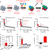
Fig. 1
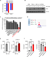
Fig. 1. Domain-specific kinome-wide CRISPR screen in high-risk ALL subtypes.
A Overview of the performed domain-specific kinome-wide library screen. SEM (KMT2A::AFF1), HAL-01 (TCF3::HLF), and TVA-1 (ETV6::ABL1) cells were virally transduced with a lentiviral Cas9-Puro construct. After puromycin selection, the cells were transduced with a GFP-tagged domain-specific kinome-wide CRISPR library consisting of 550 kinases, 100 negative controls, and 50 positive controls (6 guide RNAs (sgRNA) per kinase). Cells were collected at 2 time points for sequencing; 3 days after the library transduction, which served as input control, and 3 weeks after the transduction which served as the experimental readout. B Shown is the fold depletion of the individual sgRNAs in the tested samples. Highlighted are known positive controls for each individual ALL subtype. C Shown is the fold depletion of DYRK1A among the different ALL subtypes. D Shown is the fold depletion of each DYRK family member in SEM cells. E Cas9-puro-selected SEM cells were lentivirally transduced with a smaller sgRNA-GFP library. The library included two sgRNAs targeting one of 14 previously identified targets including 21 negative controls. Data are represented as individual values with mean ± SEM bars. *P < 0.05; **P < 0.01; ***P < 0.001 by t-test.
Fig. 2
Fig. 2. Oncogenic KMT2A rearrangements transcriptionally regulate DYRK1A in ALL.
A ChIP-Seq (Chromatin immunoprecipitation [ChIP] combined with high-throughput sequencing) tracks on the DYRK1A promoter region using antibodies specific for the KMT2A N- and the AFF1 C-terminal domain(s) were used. The Y-axis represents the number of reads for peak summit normalized by the total number of reads per track. Data from two KMT2A-AFF1 cell lines SEM (GSE83671) [34] and RS4;11 (GSE38403) [35] are shown. B ChIP-Seq tracks on the DYRK1A and MYC promoter regions using a FLAG-specific antibody in KMT2A-Aff1-FLAG-transformed human ALL cells. The Y-axis represents the number of reads for peak summit normalized by the total number of reads per track (GSE84116) [36]. C ChIP-Seq tracks on the DYRK1A and MYC promoter regions using KMT2A N-terminal-, menin-, and H3K79me2-specific antibodies. Two KMT2A-R cell lines (MOLM13/AML and RS4;11/ALL) were either treated with control or the Menin inhibitor VTP50469 as previously described (GSE127508) [10]. D Western blot analysis of DYRK1A, MYC, and β-actin in three KMT2A-R ALL cell lines (SEM, KOPN8, RS4;11) and the IgG-MYC rearranged ALL cell line MN60. Each cell line was treated with either control or the menin inhibitor VTP50469 (100 nM) for 72 h.
Fig. 3
Fig. 3. Feedback regulation between DYRK1A and MYC in KMT2A-R ALL.
A Phophoproteomics analysis in HB11;19 cells treated with 5 µM GNF2133 for 2 h and 8 h. B Western blot analysis of MYC and β-actin in KMT2A-R ALL cell lines treated either with control or EHT1610 (5 µM/72 h). C Dyrk1a gene expression levels in a comprehensive panel of purified developmentally defined normal murine B cells and genetically distinct murine lymphoma models (GSE26408) [40]. D Chip-Seq experiment on the MYC-inducible B cell malignancy model cell line P493-6. Gene tracks of Myc binding at the Dyrk1a promoter region at 0 h (top), 1 h (middle), and 24 h (bottom) are shown (GSE36354) [43]. E Myc and Dyrk1a gene expression levels in P493-6 cells before and after MYC inactivation. Myc inactivation was induced via tetracycline treatment for 48 h (GSE120246). F Myc and Dyrk1a gene expression levels in Eμ-myc transgenic mice. Data are represented as individual values with mean ± SEM bars. *P < 0.05; **P < 0.01; ***P < 0.001 by t-test.
Fig. 4
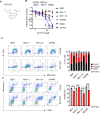
Fig. 4. DYRK1A is required for KMT2A-R ALL cell proliferation.
A Molecular structure of EHT161040. BKMT2A::AFF1 (dark blue; SEM and RS4;11), KMT2A::MLLT1 (light blue; HB11;19 and KOPN8), and non-KMT2A-R ALL cell lines (red; MN60 and Tanoue) were treated with increasing concentrations of EHT161040. Viability/cell proliferation was determined via an XTT assay after 72 h. C Cell cycle analysis of the indicated KMT2A-R ALL cell lines after treatment with 5 µM EHT1610 for 72 h. On the left are representative examples of the flow cytometric analysis (n = 3), and on the right is the summary of all three experiments. D Cell viability and apoptosis were tested in control- and DYRK1A inhibitor-treated (EHT1610; 5 µM; 72 h) KMT2A-R ALL. On the left are representative examples of the flow cytometric analysis (n = 3) and on the right is the summary of all three experiments. Data are represented as individual values with mean ± SEM bars. *P < 0.05; **P < 0.01; ***P < 0.001 by t-test.
Fig. 5
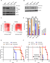
Fig. 5. DYRK1A induces cell cycle arrest via ERK signaling pathway hyperactivation.
A Protein expression levels of the indicated proteins were determined via Western blot using three KMT2A-R ALL cell lines treated with 5 µM EHT1610 for the indicated time points. B KMT2A-R (SEM and HB11;19) and Ph-like (MUTZ5 and MHH-CALL4) ALL cell lines as well as KMT2A-R (MOLM14, MV4;11) AML cell lines were treated either with control or 5 µM EHT1610. After 72 h pERK, ERK, and β-ACTIN protein levels were determined via Western blot. C The indicated KMT2A-R ALL cell lines were treated either with control, 5 µM EHT1610, 20 nM trametinib, or both drugs. After 72 h, the expression levels of the indicated proteins were determined via Western blotting. D The combinatorial effect of EHT1610 and trametinib was determined via synergy analyses using Synergy Finder software.
Fig. 6
Fig. 6. Dual DYRK1A and BCL2 inhibition synergistically kills KMT2A-R ALL.
A Western blot analysis of the indicated KMT2A-R ALL cell lines treated either with vehicle control or 5 µM EHT1610. After 72 h, the expression levels of the indicated proteins were determined. B Western blot analysis of KMT2A-R ALL cell lines treated with control or 5 µM EHT1610. After 72 h, the expression levels of BCL-XL and β-actin were determined. C SEM and KOPN8 cells were treated with increasing concentrations of EHT1610 and venetoclax. The synergistic effect of both drugs was determined via Synergy Finder software as in Fig. 5. D Flow cytometric analysis of 4 KMT2A-R ALL cell lines treated with 5 µM EHT1610 and 20 nM venetoclax for 72 h. Shown is the statistical analysis (n = 3). E Kaplan–Meier analysis of the overall survival of mice injected with 1 million KMT2A-AFF1 rearranged PDX cells (150MD) and treated either with control, GNF2133 (50 µM), venetoclax (50 µM), or a combination of both drugs. The treatment was started after ~5% CD19+/CD45+ALL cells were detected in the blood and continued until ~80% CD19+/CD45+ ALL replacements were detected in the blood of the mice. Significance was calculated via a log-rank test in Prism. F Kaplan–Meier survival analysis of KMT2A::AFF1 ALL PDX model (ALL26MD) mice treated for 20 days with vehicle control, GNF2133 (50 µM), venetoclax (50 µM), or a combination of both drugs (n = 5 mice/cohort). Significance was calculated via a log-rank test in Prism. Data are represented as individual values with mean ± SEM bars. *P < 0.05; **P < 0.01; ***P < 0.001 by t-test or Log-rank (Mantel–Cox) test.

References

    1. Tkachuk DC, Kohler S, Cleary ML. Involvement of a homolog of Drosophila trithorax by 11q23 chromosomal translocations in acute leukemias. Cell. 1992;71:691–700. - PubMed
    1. Ayton PM, Cleary ML. Molecular mechanisms of leukemogenesis mediated by MLL fusion proteins. Oncogene. 2001;20:5695–707. - PubMed
    1. Winters AC, Bernt KM. MLL-rearranged leukemias-an update on science and clinical approaches. Front Pediatr. 2017;5:4. - PMC - PubMed
    1. Schultz KR, Bowman WP, Aledo A, Slayton WB, Sather H, Devidas M, et al. Improved early event-free survival with imatinib in Philadelphia chromosome-positive acute lymphoblastic leukemia: a children’s oncology group study. J Clin Oncol. 2009;27:5175–81. - PMC - PubMed
    1. Schultz KR, Carroll A, Heerema NA, Bowman WP, Aledo A, Slayton WB, et al. Long-term follow-up of imatinib in pediatric Philadelphia chromosome-positive acute lymphoblastic leukemia: Children’s Oncology Group study AALL0031. Leukemia. 2014;28:1467–71. - PMC - PubMed

MeSH terms

Substances

LinkOut - more resources