

Clinical and spectral-domain optical coherence tomography findings and changes in new-onset macular edema after silicone oil tamponade
- PMID: 40148832
- PMCID: PMC11951619
- DOI: 10.1186/s12886-025-03986-0
Clinical and spectral-domain optical coherence tomography findings and changes in new-onset macular edema after silicone oil tamponade
Abstract
Backgroud: Few investigations have been conducted on the detailed clinical features of CME associated with SiO, from emergence to restoration, especially using OCT images. This study aimed to analyze the clinical and spectral-domain optical coherence tomography (SD-OCT) characteristics and changes in cystoid macular edema (CME) associated with silicone oil (SiO).
Methods: Retrospective case series. Six cases of newly on-set CME after SiO tamponade were examined. SD-OCT was performed before pars plana vitrectomy, after SiO tamponade, and after SiO removal. Clinical and SD-OCT data was collected.
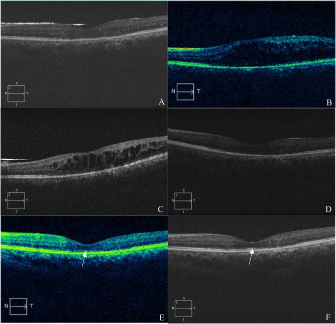
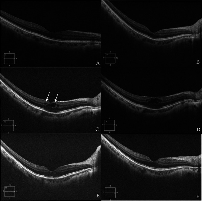
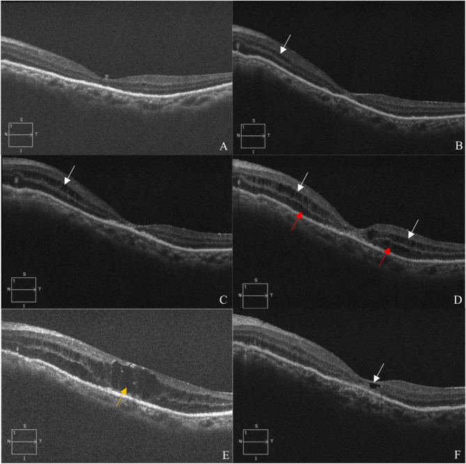
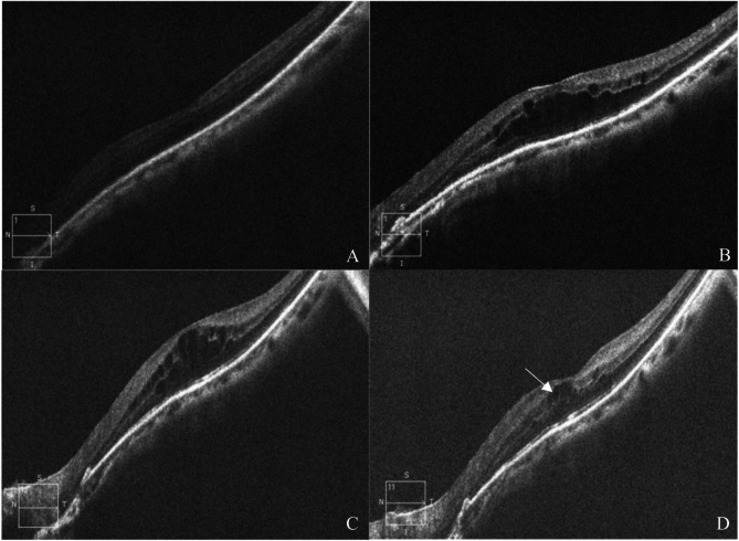
Results: CME was first noted at 28.83 ± 9.22 days after SiO tamponade. The average foveal thickness was 411 ± 41 μm before oil removal and decreased to 267 ± 69 μm three days after oil removal (P = 0.028). The average visual acuity before and after oil removal were 0.82 ± 0.40 logarithm of the minimum angle of resolution (logMAR) and 0.75 ± 0.45 logMAR, respectively, and the difference was not statistically significant (P = 0.285). SD-OCT revealed that three patients had edema first in the inner nuclear layer (INL), and three had cysts in both INL and outer nuclear layers (ONL) at discovery. Of the six patients, three exhibited cystic changes in the fovea firstly. CME showed rapid recovery following SiO removal, with cysts completely disappearing in four patients (66.7%) within 3 days. However, in two patients (33.3%), the cysts persisted in INL after three days, whereas the cysts in ONL had resolved completely. The ellipsoid zone integrity of the macular region was smoother in patient with better vision.
Conclusion: New-onset CME after SiO tamponade may initially affects INL and then ONL. CME shows significant improvement after oil removal, probably initially resolving in ONL, and then followed by INL. SD-OCT enabless monitoring of macular microstructure changes in SiO-treated eyes, and macular cysts' occurrence can indicate the oil removal need.
Keywords: Cystoid macular edema; Silicone oil eyes; Silicone oil removal; Spectral domain optical coherence tomography.
© 2025. The Author(s).
Conflict of interest statement
Declarations. Ethics approval and consent to participate: We adhered to the tenets of the Declaration of Helsinki. Ethics approval was obtained from the the ethics committee of Beijing Tongren hospital. All participants involved were informed of the purpose of this study and an informed consent was obtained from them. Consent for publication: Not Applicable. Competing interests: The authors declare no competing interests.
Figures
References
-
- Daruich A, Matet A, Moulin A, Kowalczuk L, Nicolas M, Sellam A, Rothschild PR, Omri S, Gélizé E, Jonet L, et al. Mechanisms of macular edema: beyond the surface. Prog Retin Eye Res. 2018;63:20–68. - PubMed
-
- Spaide RF. Retinal vascular cystoid macular edema: review and new theory. Retina. 2016;36(10):1823–42. - PubMed
-
- Shalchi Z, Mahroo OA, Shunmugam M, Mohamed M, Sullivan PM, Williamson TH. Spectral domain optical coherence tomography findings in long-term silicone oil-related visual loss. Retina. 2015;35(3):555–63. - PubMed
-
- Lo DM, Flaxel CJ, Fawzi AA. Macular effects of silicone oil tamponade: optical coherence tomography findings during and after silicone oil removal. Curr Eye Res. 2017;42(1):98–103. - PubMed
MeSH terms
Substances
LinkOut - more resources
Full Text Sources
Medical
Miscellaneous
