. 2025 Feb 25;15(3):333.
doi: 10.3390/biom15030333.

Inhibition of DPP-4 Attenuates Endotoxemia-Induced NLRC4 Inflammasome and Inflammation in Visceral Adipose Tissue of Mice Fed a High-Fat Diet

Affiliations

Inhibition of DPP-4 Attenuates Endotoxemia-Induced NLRC4 Inflammasome and Inflammation in Visceral Adipose Tissue of Mice Fed a High-Fat Diet

Francesca Bianchi et al. Biomolecules. .

Abstract

Inflammasomes are protein complexes that trigger pro-inflammatory responses and promote many diseases, including adipose tissue dysfunction. Linagliptin (L), a DPP-4 inhibitor used for type 2 diabetes therapy, has putative anti-inflammatory effects. This work explores L effects on inflammasome regulation, inflammation, and adipose tissue dysfunction in obese mice. Male C57BL/6N mice were fed a normal chow (NC) diet, high-fat (HF) diet, or HF diet with L (HFL) for 15 weeks. Gene expression and histological examinations were performed on visceral (VAT) and subcutaneous (SAT) adipose tissue samples. Biomarkers were quantified on sera. Murine macrophages were utilized for in vitro analyses. L decreased HF-induced endotoxemia and circulating inflammatory indicators. Despite having no effect on body weight, L reduced VAT inflammation by decreasing endotoxemia-induced NLRC4 inflammasome, inflammation severity, and fat cell hypertrophy. Although SAT response differed from VAT, inflammation was slightly reduced in this tissue too. In vitro, L modulated inflammation by directly reducing the pro-inflammatory macrophage phenotype. In obesity, increased NLRC4 inflammasome expression links endotoxemia and VAT inflammation. L protected against endotoxemia, maybe by affecting gut permeability and VAT responses. The decreased polarization of macrophages toward a pro-inflammatory phenotype and the reduction in adipocyte hypertrophy are involved in the response to L.

Keywords: DPP-4; NLRC-4 inflammasome; adipocytes; endotoxemia; fat; high-fat diet; inflammasomes; inflammation; linagliptin; macrophage phenotype.


Conflict of interest statement

The authors have no conflicts of interest to declare.

Figures

Figure 1
Figure 1
Effect of high-fat diet (HF) and high-fat diet combined with Linagliptin (HFL) on body weight. The figure shows the changes in body weight across the experimental weeks in each group. a, p < 0.001 vs. week 0; b, p < 0.01 vs. week 7; c, p < 0.001 vs. week 7; * p < 0.05 vs. NC, *** p < 0.001 vs. NC. NC, normal chow; HF, high fat; HFL, high-fat Linagliptin.
Figure 2
Figure 2
Effect of high-fat diet (HF) and high-fat diet combined with Linagliptin (HFL) on serum level of lipopolysaccharide binding protein (LBP). The figure shows the effect of HF diet and HFL diet on serum LBP concentration. ** p < 0.01, *** p < 0.001.
Figure 3
Figure 3
Histological analysis of visceral adipose tissue (VAT) and subcutaneous adipose tissue (SAT) of the three experimental groups (NC, normal chow; HF, high fat; FHL, high fat with Linagliptin). Microscopical morphology of VAT (panel AC) and SAT (panel DF) adipocytes stained with hematoxylin and eosin. VAT and SAT adipocytes of mice fed NC diet have a normal size with variably evident mostly plump nuclei (panel A,D), while those of mice fed HF diet are 2–4 times larger than NC group (panel B,E). In HFL group (panel C,F), adipocytes have similar morphology of those in HF group. Images are at 400× magnification. Bars = 50 microns.
Figure 4
Figure 4
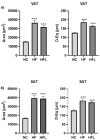
Effect of high-fat diet (HF) and high-fat diet combined with Linagliptin (HFL) on adipocyte equivalent diameter (D-Eq) and area. The figure shows the effect of HF diet and HFL diet on D-Eq and area of visceral (VAT, panel A) and subcutaneous (SAT, panel B) adipocytes. *** p < 0.001 vs. NC. In (panel A), the difference between HFL and HF was at limit of statistical significance both for area (p = 0.07) and D-Eq (p = 0.051).
Figure 5
Figure 5
Effect of high-fat diet (HF) and high-fat diet combined with Linagliptin (HFL) on macrophage polarization in vivo. The figure shows the effect of HF and HFL diet on the expression of markers associated to M1- and M2-phenotype macrophage polarization in visceral adipose tissue. Data are expressed as 2^(-ΔΔCt) vs. NC (normal chow; dotted line). CD68 (cluster differentiation 68, panel A) is an overall marker of macrophage, ITAGX (Integrin Subunit Alpha X, panel B) is a marker of M1-polarized macrophages, and SLC2A4 encodes glucose transporter Type 4 (Solute Carrier Family 2 Member 4, panel C). IL-10 (Interelukin-10, panel D) and MRC1 (Mannose Receptor C-Type 1, panel E) are markers of M2-polarized macrophages. * p < 0.05 vs. NC, ** p < 0.01 vs. NC, ° p < 0.05 vs. HF.
Figure 6
Figure 6
Effect Linagliptin (L) on macrophage polarization in vitro. The figure shows the effects of two L doses (100 and 500 nM) on the expression of markers associated to M1-phenotype macrophage polarization in vitro at 6 h and 24 h. Data are expressed as 2^(-ΔΔCt) vs. M0 and M1-polarized macrophages (dotted lines). ITAGX (Integrin Subunit Alpha X, panel A) and NOS2 (Nitric Oxide Synthase 2, panel B) are markers of M1-polarized macrophages.
Figure 7
Figure 7
Effect of high-fat diet (HF) and high-fat diet combined with Linagliptin (HFL) on circulating chemokines and cytokines. (AE) shows the effect of HF diet and HFL diet on plasma levels of different cytokines and chemokines. * p < 0.05, ** p < 0.01, and *** p < 0.001. CCL11, C-C Motif Chemokine Ligand 11; CCL7, chemokine CC motif ligand 7; IL-33, interleukin-33; DPP-4; IL-17A, interleukin-17A.

References

    1. Eckel R.H., Grundy S.M., Zimmet P.Z. The metabolic syndrome. Lancet. 2005;365:1415–1428. doi: 10.1016/S0140-6736(05)66378-7. - DOI - PubMed
    1. Vianello E., Dozio E., Arnaboldi F., Marazzi M.G., Martinelli C., Lamont J., Tacchini L., Sigruner A., Schmitz G., Corsi Romanelli M.M. Epicardial adipocyte hypertrophy: Association with M1-polarization and toll-like receptor pathways in coronary artery disease patients. Nutr. Metab. Cardiovasc. Dis. 2016;26:246–253. doi: 10.1016/j.numecd.2015.12.005. - DOI - PubMed
    1. Cinti S., Mitchell G., Barbatelli G., Murano I., Ceresi E., Faloia E., Wang S., Fortier M., Greenberg A.S., Obin M.S. Adipocyte death defines macrophage localization and function in adipose tissue of obese mice and humans. J. Lipid Res. 2005;46:2347–2355. doi: 10.1194/jlr.M500294-JLR200. - DOI - PubMed
    1. Cani P.D., Amar J., Iglesias M.A., Poggi M., Knauf C., Bastelica D., Neyrinck A.M., Fava F., Tuohy K.M., Chabo C., et al. Metabolic endotoxemia initiates obesity and insulin resistance. Diabetes. 2007;56:1761–1772. doi: 10.2337/db06-1491. - DOI - PubMed
    1. Halberg N., Khan T., Trujillo M.E., Wernstedt-Asterholm I., Attie A.D., Sherwani S., Wang Z.V., Landskroner-Eiger S., Dineen S., Magalang U.J., et al. Hypoxia-inducible factor 1alpha induces fibrosis and insulin resistance in white adipose tissue. Mol. Cell Biol. 2009;29:4467–4483. doi: 10.1128/MCB.00192-09. - DOI - PMC - PubMed

MeSH terms

LinkOut - more resources