

The Human Thyroid-Derived CI-huThyrEC Cell Line Expresses the Thyrotropin (TSH) Receptor and Thyroglobulin but Lacks Other Essential Characteristics of Thyroid Follicular Cells
- PMID: 40149910
- PMCID: PMC11940677
- DOI: 10.3390/biom15030375
The Human Thyroid-Derived CI-huThyrEC Cell Line Expresses the Thyrotropin (TSH) Receptor and Thyroglobulin but Lacks Other Essential Characteristics of Thyroid Follicular Cells
Abstract
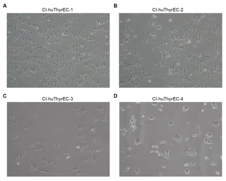
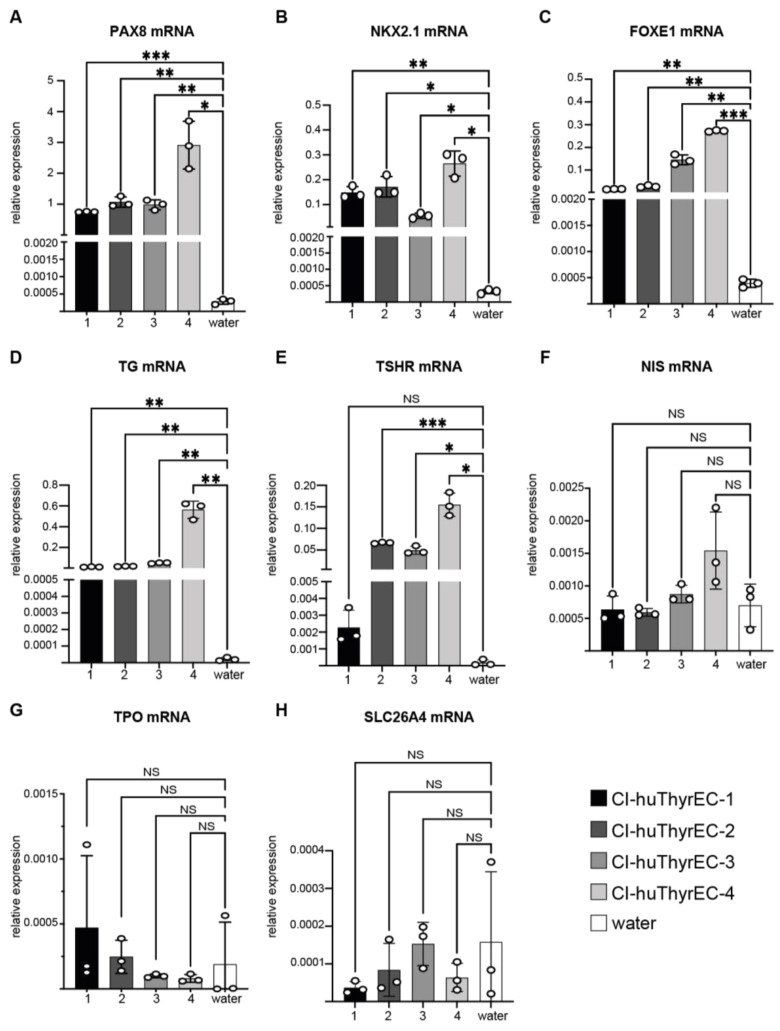
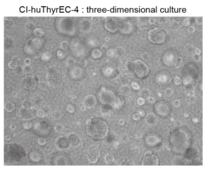
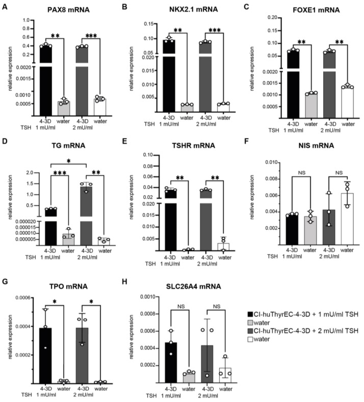
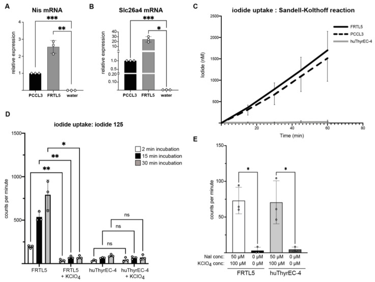
Background: Thyroid hormone synthesis requires the normal function of thyroid follicular cells and adequate nutritional intake of iodine. For in vitro studies on thyroid cell pathophysiology, the immortalized FRTL5 rat thyroid cell line and a derivative thereof, the PCCL3 cell line, are widely used. However, a permanent human thyroid cell line is currently lacking. A recent report described a cell line obtained from human thyroid cells designated as Cl-huThyrEC. Methods: Four clones of Cl-huThyrEC cells were obtained and cultured in the presence of thyroid stimulating hormone (TSH). The expression of key genes defining the thyroid follicular cell phenotype was determined by reverse-transcription PCR (RT-PCR) in FRTL5, PCCL3, and Cl-huThyrEC cells. The latter were cultured as monolayers and as organoids in Matrigel. Iodide uptake was measured and compared among the cell lines. Results: Gene expression analysis reveals that Cl-huThyrEC cells express the thyroid-restricted transcription factors (PAX8, NKX2.1, FOXE1), the TSH receptor (TSHR), and thyroglobulin (TG), but they do not express the sodium-iodide symporter (NIS), thyroid peroxidase (TPO), and pendrin (SLC26A4). In functional studies, Cl-huThyrEC cells are unable to concentrate iodide. Conclusions: Despite the expression of certain key genes that are limited or restricted to thyroid follicular cells, Cl-huThyrEC cells lack some of the essential characteristics of thyroid follicular cells, in particular, NIS. Hence, their utility as a model system for thyroid follicular cells is limited.
Keywords: cell line; iodine; sodium iodide symporter; thyroid; thyroid peroxidase.
Conflict of interest statement
The authors declare no conflicts of interest. The funders had no role in the design of the study; in the collection, analyses, or interpretation of data; in the writing of the manuscript; or in the decision to publish the results.
Figures

Similar articles
-
Thyroid follicle formation and thyroglobulin expression in multipotent endodermal stem cells.Thyroid. 2013 Apr;23(4):385-91. doi: 10.1089/thy.2012.0644. Epub 2013 Mar 18. Thyroid. 2013. PMID: 23360087 Free PMC article.
-
Thyrotropin regulation of differentiated gene transcription in adult human thyrocytes in primary culture.Mol Cell Endocrinol. 2020 Dec 1;518:111032. doi: 10.1016/j.mce.2020.111032. Epub 2020 Sep 14. Mol Cell Endocrinol. 2020. PMID: 32941925 Free PMC article.
-
TAZ Induction Directs Differentiation of Thyroid Follicular Cells from Human Embryonic Stem Cells.Thyroid. 2017 Feb;27(2):292-299. doi: 10.1089/thy.2016.0264. Epub 2017 Jan 3. Thyroid. 2017. PMID: 27829313 Free PMC article.
-
Iodide handling disorders (NIS, TPO, TG, IYD).Best Pract Res Clin Endocrinol Metab. 2017 Mar;31(2):195-212. doi: 10.1016/j.beem.2017.03.006. Epub 2017 Apr 4. Best Pract Res Clin Endocrinol Metab. 2017. PMID: 28648508 Review.
-
Thyroglobulin regulates follicular function and heterogeneity by suppressing thyroid-specific gene expression.Biochimie. 1999 Apr;81(4):329-40. doi: 10.1016/s0300-9084(99)80078-9. Biochimie. 1999. PMID: 10401666 Review.
References
-
- Kopp P. Thyroid hormone synthesis. In: Braverman L.E., Cooper D.S., Kopp P., editors. Werner and Ingbar’s the Thyroid: A Fundamental and Clinical Text. Wolters Kluwer; Philadelphia, PA, USA: 2021. pp. 59–85.
-
- Bianco A.C., Anderson G., Forrest D., Galton V.A., Gereben B., Kim B.W., Kopp P.A., Liao X.H., Obregon M.J., Peeters R.P., et al. American Thyroid Association Guide to investigating thyroid hormone economy and action in rodent and cell models. Thyroid. 2014;24:88–168. doi: 10.1089/thy.2013.0109. - DOI - PMC - PubMed
MeSH terms
Substances
Grants and funding
LinkOut - more resources
Full Text Sources
Miscellaneous
