. 2025 Mar 12;15(3):406.
doi: 10.3390/biom15030406.

Electrostatic Effects on Tau Nanocondensates

Affiliations

Electrostatic Effects on Tau Nanocondensates

Phoebe S Tsoi et al. Biomolecules. .

Abstract

Biomolecular condensates (BMCs) are membrane-less protein compartments with physiological and pathological relevance. The formation of BMCs is driven by a process known as liquid-liquid phase separation (LLPS), a field that has largely focused on the study of micron-sized condensates. However, there have been recent studies showing that proteins that undergo LLPS also form nanometer-sized condensates. These nanometer-sized condensates, or nanocondensates, are distinct from microcondensates and potentially exhibit more relevance in cell biology. The field of nanocondensate research is in its infancy, with limited biophysical studies of these structures. Here, we studied condensate formation and dissolution of wild-type and disease-linked (hyperphosphorylated and missense mutated) Tau. We investigated the effects of solution condition modulation on nanocondensate formation and dissolution, and observed that Tau condensation is strongly regulated by electrostatic forces and less affected by hydrophobic disruption. We observed that all three Tau variants studied shared condensate formation properties when in solution conditions with the same ionic strength. However, hyperphosphorylated and missense-mutated Tau exhibited higher resistance to dissolution compared to wild-type Tau. This study uncovers additional distinctions between different types of condensates, which provides further insight into the distinctions between physiological and pathological condensates.

Keywords: LLPS; Tau; biomolecular condensates; nanocondensates; neurodegeneration; protein condensation; tauopathy.


Conflict of interest statement

The authors declare no conflicts of interest.

Figures

Figure 1
Figure 1
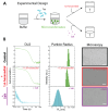
Phase separation of Tau monitored by dynamic light scattering (DLS). (A) Domain organizations and charge distributions of wild-type and P301S Tau compared to hyperphosphorylated Tau (pTau), highlighting differences in isoelectric points (pH 8.2 vs. 4.7), and displaying mutation and post-translational modification sites. Tau consists of two N-terminal inserts (N1, N1), three proline-rich domains (P1–P3), and four microtubule-binding repeats (R1–R4). (B) Tau can exist as monomers, nanocondensates, and microcondensates, detected using DLS and optical microscopy. Representative data from wild-type Tau are shown. Solution conditions: 20 μM Tau in water (monomers, 2–10 nm); 1μM Tau in 20 mM HEPES buffer, pH 8 (nanocondensates, 80–300 nm); and 20 μM Tau in 20 mM HEPES buffer, pH 8 (microcondensates, > 700 nm). Scale bars represent 10 μm.
Figure 2
Figure 2
WT Tau microcondensates are modulated by salt, but unperturbed by hexanediol. (A) Schematic of salt and hexanediol modulation of Tau microcondensates. (B) A total of 20 μM WT Tau in 20 mM HEPES, pH 8, forms microcondensates detected by DLS and optical microscopy. The addition of 5% hexanediol does not significantly affect Tau condensation while the addition of 200 mM NaCl reverses condensates into the monomeric state. Scale bars represent 10 μm.
Figure 3
Figure 3
Formation and dissolution of WT Tau and pTau nanocondensates, modulated by salt. (A) Schematic of formation and dissolution of Tau condensates via salt modulation. (B) Diagram of WT Tau condensate formation (top) and dissolution (bottom) in the presence of salt. (C) Diagram of pTau condensate formation (top) and dissolution (bottom) in the presence of salt.
Figure 4
Figure 4
Formation and dissolution of P301S Tau nanocondensates, modulated by salt. (A) P301S condensate formation (top) and dissolution (bottom) in the presence of salt. (B) Autoattenuation and peak populations observed via DLS reveal monomer (blue), multimodal (blue to orange), and nanocondensate (orange) populations of P301S Tau.

References

    1. Barbier P., Zejneli O., Martinho M., Lasorsa A., Belle V., Smet-Nocca C., Tsvetkov P.O., Devred F., Landrieu I. Role of Tau as a Microtubule-Associated Protein: Structural and Functional Aspects. Front. Aging Neurosci. 2019;11:204. doi: 10.3389/fnagi.2019.00204. - DOI - PMC - PubMed
    1. Gyparaki M.T., Arab A., Sorokina E.M., Santiago-Ruiz A.N., Bohrer C.H., Xiao J., Lakadamyali M. Tau forms oligomeric complexes on microtubules that are distinct from tau aggregates. Proc. Natl. Acad. Sci. USA. 2021;118:e2021461118. doi: 10.1073/pnas.2021461118. - DOI - PMC - PubMed
    1. Kosik K.S., Han S. Tau Condensates. Adv. Exp. Med. Biol. 2019;1184:327–339. doi: 10.1007/978-981-32-9358-8_24. - DOI - PubMed
    1. Longfield S.F., Mollazade M., Wallis T.P., Gormal R.S., Joensuu M., Wark J.R., van Waardenberg A.J., Small C., Graham M.E., Meunier F.A., et al. Tau forms synaptic nano-biomolecular condensates controlling the dynamic clustering of recycling synaptic vesicles. Nat. Commun. 2023;14:7277. doi: 10.1038/s41467-023-43130-4. - DOI - PMC - PubMed
    1. Siahaan V., Tan R., Humhalova T., Libusova L., Lacey S.E., Tan T., Dacy M., Ori-McKenney K.M., McKenney R.J., Braun M., et al. Microtubule lattice spacing governs cohesive envelope formation of tau family proteins. Nat. Chem. Biol. 2022;18:1224–1235. doi: 10.1038/s41589-022-01096-2. - DOI - PMC - PubMed

LinkOut - more resources