. 2025 Apr;45(2):311-325.
doi: 10.19852/j.cnki.jtcm.2025.02.006.

Xiahuo Pingwei San attenuated intestinal inflammation in dextran sulfate sodium-induced ulcerative colitis mice through inhibiting the receptor for advanced glycation end-products signaling pathway

Affiliations

Xiahuo Pingwei San attenuated intestinal inflammation in dextran sulfate sodium-induced ulcerative colitis mice through inhibiting the receptor for advanced glycation end-products signaling pathway

Huang Jiaen et al. J Tradit Chin Med. 2025 Apr.

Abstract

Objective: To evaluate the therapeutic effects of Xiahuo Pingwei San (, XHPWS) on ulcerative colitis (UC) in mice and to explore the underlying mechanisms through a network pharmacology approach.

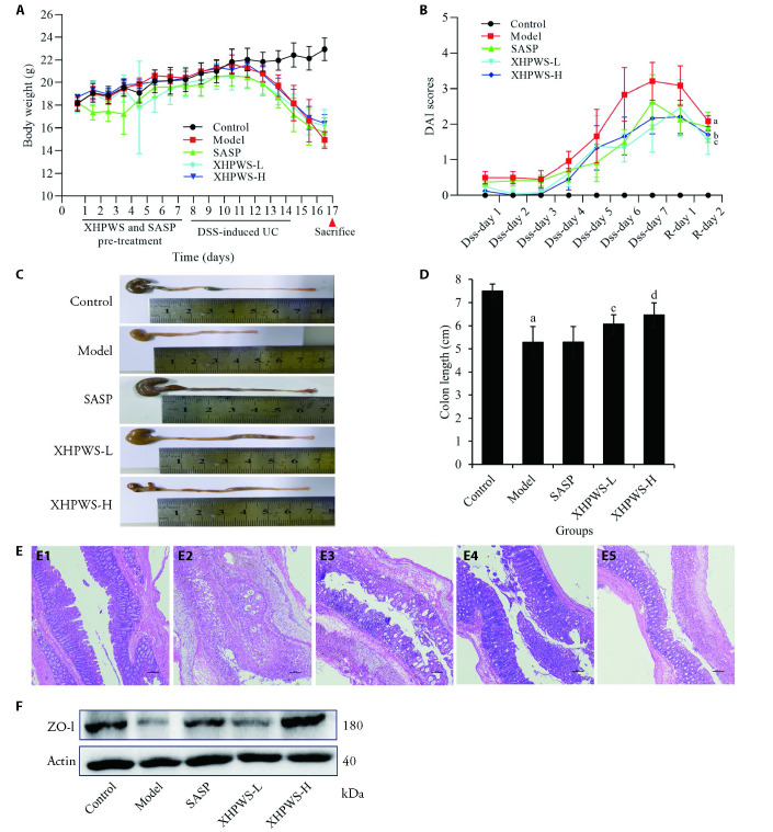
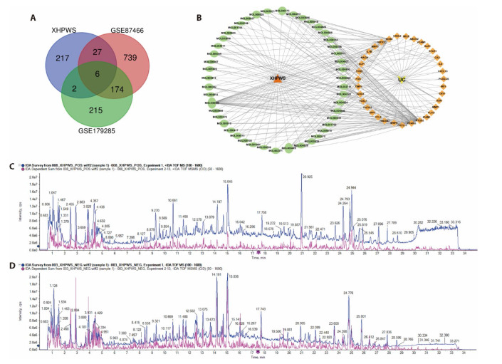
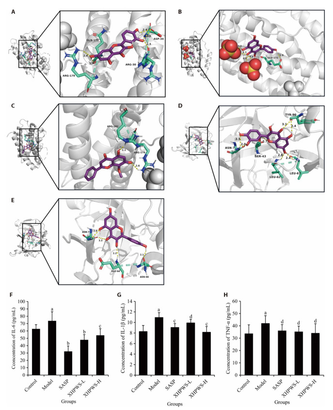
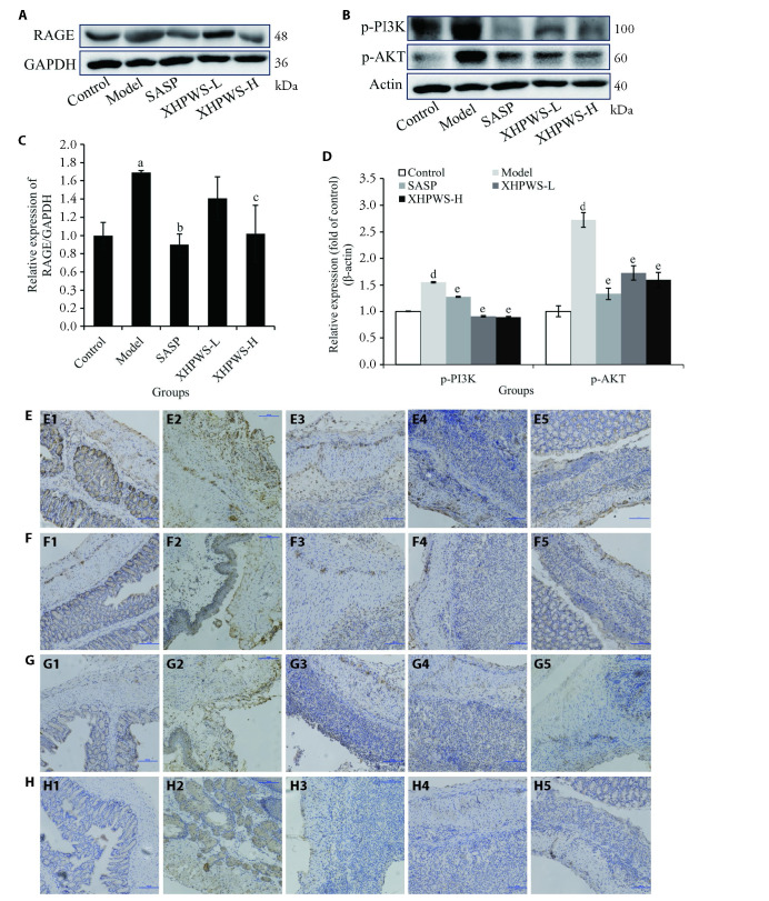
Methods: Ultra-performance liquid chromatography coupled with quadrupole time-of-flight mass spectrometry (UPLC-Q-TOF/MS) was utilized to identify the chemical composition and authenticate the active constituents of XHPWS, ensuring rigorous quality control across batches. A dextran sulfate sodium (DSS)-induced UC model was established in C57BL/6 mice, which were treated with XHPWS in vivo. The efficacy against UC was assessed by measuring parameters such as body weight, disease activity index (DAI) scores, and colon length. Levels of inflammatory cytokines, including interleukin-6 (IL-6), interleukin-1β (IL-1β), and tumor necrosis factor-alpha (TNF-α), in colonic tissue were evaluated using enzyme-linked immunosorbent assay (ELISA). Histological analysis of colon sections was conducted using hematoxylin and eosin staining. A network pharmacology approach was employed to explore the mechanisms of XHPWS and to predict its potential targets in UC treatment. Predicted protein expressions in colonic tissue were validated using immune-ohistochemistry (IHC) and Western blotting techniques.

Results: XHPWS effectively alle viated DSS-induced UC symptoms in mice, as evidenced by restored body weight, reduced colon shortening, and decreased DAI scores. Histopathological examination revealed that XHPWS significantly reduced intestinal inflammatory infiltration, restored intestinal epithelial permeability, and increased goblet cell count. Network pharmacology analysis identified 63 active compounds in XHPWS and suggested that it might target 35 potential proteins associated with UC treatment. Functional enrichment analysis indicated that the protective mechanism of XHPWS could be related to the advanced glycation end products-receptor for advanced glycation end products (AGE-RAGE) signaling pathway. Notably, quercetin, kaempferol, wogonin, and nobiletin, the main components of XHPWS, showed strong correlations with the core targets. Additionally, experimental validation demonstrated that XHPWS significantly decreased levels of inflammatory cytokines interleukin 6 (IL-6), interleukin 1 beta (IL-1β), and tumor necrosis factor alpha (TNF-α) in UC mice, while downregulating the expression of proteins related to the AGE-RAGE pathway.

Conclusion: Our study demonstrated that XHPWS effectively alle viates colitis symptoms and inflammation in UC mice, potentially through the regulation of the AGE-RAGE pathway. These findings provide strong evidence for the therapeutic potential of XHPWS in UC treatment, thereby broadening its clinical applications.

Keywords: Xiahuo Pingwei San; colitis, ulcerative; glycation end products, advanced; inflammation; network pharmacology; receptor for advanced glycation end products; signal transduction.


Figures

Figure 1
Figure 1. XHPWS alleviated symptoms of UC mice induced by DSS
A: the animal study design and the time-course of body weight changes; B: the time-course of disease activity index in different group; C: representative colon images in each group. D: the colon length in each group. E: Histological analysis of colon tissues by HE staining (× 100, scale bars = 100 μm); E1: colon tissues of the control group; E2: colon tissues of the model group; E3: colon tissues of the SASP group; E4: colon tissues of the XHPWS-L group; E5: colon tissues of the XHPWS-H group. F: representative Western blotting brands of ZO-1 in the colon tissues. Control: normal control (fed on standard chow); Model: model group (DSS + standard chow); SASP: positive control group (DSS + 200 mg/kg SASP); XHPWS-L: low-dose XHPWS group (DSS + 5.46 g/kg XHPWS); XHPWS-H: high-dose XHPWS group (DSS + 10.92 g/kg XHPWS). XHPWS: Xiahuo Pingwei San; DSS: dextran sulfate sodium; SASP: sulfasalazine; ZO-1: Zonula Occludens 1; UC: ulcerative colitis; HE: hematoxylin and eosin; ANOVA: analysis of variance. Statistical significance was assessed using one-way ANOVA, followed by pairwise comparisons between groups using the t-test. All data were expressed as the mean ± standard deviation (n = 8). aP < 0.01, compared with the control group; bP < 0.001, cP < 0.05 and dP < 0.01, compared with the model group.
Figure 2
Figure 2. Active Ingredients-Targets network of XHPWS and potential target genes
A: venn diagram of the targets both in the differential genes of UC and XHPWS targets; B: active ingredients-targets network, the edges represented the relationship between active ingredients and the target genes; C: positive ion mode UPLC-Q-TOF/MS base peak ion flow graph (BPC) for XHPWS; D: negative ion mode UPLC-Q-TOF/MS BPC for XHPWS. XHPWS: Xiahuo Pingwei San; UC: ulcerative colitis; UPLC-Q-TOF/MS: ultra-performance liquid chromatography coupled with quadrupole time-of-flight mass spectrometry.
Figure 3
Figure 3. Molecular docking simulation for the compounds of HXPWS binding with IL-6 and IL-1β
Molecular docking simulation between molecular compounds and proteins: A: quercetin and IL-6; B: kaempferol and IL-6; C: wogonin and IL-6; D: quercetin and IL-1β; E: kaempferol and IL-1β. Effects of XHPWS on pro-inflammatory cytokines F: IL-6, G: IL-1β, and H: TNF-α in colonic tissues of UC mice determined by ELISA assay. Control: normal control (fed on standard chow); Model: model group (DSS + standard chow); SASP: positive control group (DSS + 200 mg/kg SASP); XHPWS-L: low-dose XHPWS group (DSS + 5.46 g/kg XHPWS); XHPWS-H: high-dose XHPWS group (DSS + 10.92 g/kg XHPWS). XHPWS: Xiahuo Pingwei San; DSS: dextran sulfate sodium; SASP: sulfasalazine; UC: ulcerative colitis; ANOVA: analysis of variance; ELISA: enzyme-linked immunosorbent assay; IL-6: interleukin-6; IL-1β: interleukin-1β; TNF-α: tumor necrosis factor-alpha. Statistical significance was assessed using one-way ANOVA, followed by pairwise comparisons between groups using the t-test. All data were expressed as the mean ± standard deviation (n = 8). aP < 0.05, compared with the control group; bP < 0.001, cP < 0.01 and dP < 0.05, compared with the model group.
Figure 4
Figure 4. Modulation of the expression of RAGE and its downstream related proteins by XHPWS
A: the expressions of RAGE in colon tissues of mice were evaluated by Western blot; B: the expressions of p-PI3K, p-AKT in colon tissues of mice were evaluated by Western blot; C: quantitative analysis of RAGE for western blot results by normalizing to GAPDH; D: quantitative analysis of p-PI3K and p-AKT for Western blot results by normalizing to β-actin; E: representative images of PI3K immunostaining of colon tissues in each group (× 200, bar = 100 μm); F: representative images of AKT immunostaining of colon tissues in each group (× 200, bar = 100 μm); G: representative images of NF-κB p65 immunostaining of colon tissues in each group (× 200, bar = 100 μm); H: representative images of NF-κB p-p65 immunostaining of colon tissues in each group (× 200, bar = 100 μm); E1, F1, G1, H1: colon tissues of the control group; E2, F2, G2, H2: colon tissues of the model group; E3, F3, G3, H3: colon tissues of the SASP group; E4, F4, G4, H4: colon tissues of the XHPWS-L group; E5, F5, G5, H5: colon tissues of the XHPWS-H group. Control: normal control (fed on standard chow); Model: model group (DSS + standard chow); SASP: positive control group (DSS + 200 mg/kg SASP); XHPWS-L: low-dose XHPWS group (DSS + 5.46 g/kg XHPWS); XHPWS-H: high-dose XHPWS group (DSS + 10.92 g/kg XHPWS). XHPWS: Xiahuo Pingwei San; DSS: dextran sulfate sodium; SASP: sulfasalazine; RAGE: receptor for advanced glycation end products; PI3K: phosphatidylinositol 3-kinase; AKT: protein kinase B; NF-κB p65: nuclear factor kappa B p65 subunit; GAPDH: glyceraldehyde-3-phosphate dehydrogenase; ANOVA: analysis of variance. Statistical significance was assessed using one-way ANOVA, followed by pairwise comparisons between groups using the t-test. All data are expressed as the mean ± standard deviation (n = 3). aP < 0.01 and dP < 0.001, compared with the control group; bP < 0.01, cP < 0.05 and eP < 0.001, compared with the model group.

Similar articles

References

    1. Kirsner JB. Historical origins of current IBD concepts. World J Gastroenterol 2001; 7: 175-84. - PMC - PubMed
    1. Kaplan GG. The global burden of IBD: from 2015 to 2025. Nat Rev Gastroenterol Hepatol 2015; 12: 720-7. - PubMed
    1. Yao D, Dong M, Dai C, Wu S. Inflammation and inflammatory cytokine contribute to the initiation and development of ulcerative colitis and its associated cancer. Inflamm Bowel Dis 2019; 25: 1595-602. - PubMed
    1. Rubio CA, Langner C, Schmidt PT. Partial to complete abrogation of the subepithelial macrophage barrier against the gut microbiota in patients with ulcerative colitis and Crohn's colitis. Histopathology 2018; 72: 580-97. - PubMed
    1. Lawrence T, Natoli G. Transcriptional regulation of macrophage polarization: enabling diversity with identity. Nat Rev Immunol 2011; 11: 750-61. - PubMed

MeSH terms

LinkOut - more resources