. 2025 Aug;247(2):393-407.
doi: 10.1111/joa.14251. Epub 2025 Mar 28.

Topohistology of dendritic cells and macrophages in the distal and proximal nodes along the lymph flow from the lung

Affiliations

Topohistology of dendritic cells and macrophages in the distal and proximal nodes along the lymph flow from the lung

Masaya Aoki et al. J Anat. 2025 Aug.

Abstract

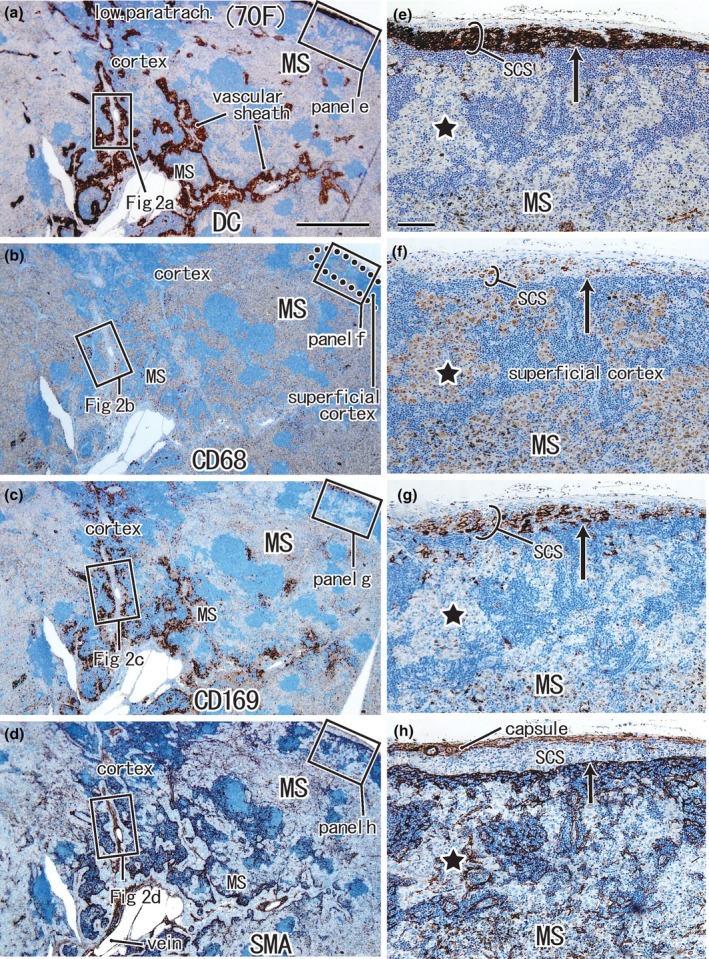
Nodal dendritic cells and CD169-positive macrophages cross-present cancer antigens earlier in the proximal nodes than in the distal nodes along the lymph flow from cancer. We examined topohistological differences between the proximal and distal nodes before the formation of metastasis. Immunohistochemical and morphometric analyses were performed to examine DC-SIGN-, CD68-, and CD169-positive cells in the subcarinal node (proximal) and paratracheal nodes (distal nodes) from 16 patients with lower-lobe lung cancer without metastasis (adenocarcinoma, 11; squamous, 5). Nodes at the same sites from 10 patients with upper-lobe cancer were used as controls. In all nodes, the medullary sinus was filled with CD68-positive and CD169-negative macrophages, most of which showed anthracosis. The proximal node carried a significantly smaller overlap between clusters of DC-SIGN-positive cells and CD169-positive cells relative to the distal node in lower-lobe cancer patients (p = 0.015). Irrespective of the cancer pathology, the tumor size was significantly correlated with the longer subcapsular clusters containing either DC-SIGN-positive cells or CD169-positive cells (p = 0.003, 0.043). A significantly small overlap between these clusters as well as the missing paracortical sinuses was evident in the negative control node outside the lymph flow (p = 0.006). Since DC-SIGN-positive cells and CD169-positive cells, especially composite cells in the overlapped cluster, are likely to be derived from monocytes, larger tumors appeared to accelerate the migration into the subcapsular sinus. In contrast to the suggested active status of the distal node, the proximal node appeared to have already been suppressed. This downregulation reached the level in the negative control node.

Keywords: CD169‐positive macrophages; anti‐cancer immunity; dendritic cells; lung regional node; morphometry; preconditioning along lymph flow; tumor size.


Conflict of interest statement

The authors declare no conflicts of interest in association with the present study.

Figures

FIGURE 1
FIGURE 1
Positive control node (lower paratracheal node) from a 70‐year‐old female patient with upper‐lobe lung cancer. Panels (a)–(d) display adjacent or near sections. Panels (e)–(h) show higher magnification views of the squares in panels (a)–(d), respectively. Immunohistochemistry for DCsign (panels a and e), CD68 (panels b and f), CD169 (panels c and g), and smooth muscle actin or SMA (panels d and h). DCsign‐positive cells are observed in the subcapsular sinus (SCS) and along the vascular sheath (panels a and e). A higher magnification of the vascular sheath (squares with figure number in panels a–d) is shown in Figure 2. The subcapsular sinus (SCS) is filled with DCsign‐positive cells and CD169‐positive cells, but it contains few CD68‐positive macrophages (long arrow in panels e–g). The nodal capsule and endothelium of the sinuses express SMA (panel h). SMA‐positive endothelia are also observed in lymphocyte clusters (i.e., the cortex). The medullary sinus (MS) inserts into the superficial cortex (star in panels e–g) and contains abundant CD68‐positive and CD169‐negative macrophages. Panels (a)–(d) or panels (e)–(h) were prepared at the same magnification (scale bars, 1 mm in panel a; 0.1 mm in panel e).
FIGURE 2
FIGURE 2
Higher magnification views of DCsign‐positive cells and macrophage clusters in the medullary sinus and cortex (same specimen as Figure 1). Panels (a)–(d) correspond to squares in Figure 1(a)–(d), but the up‐down orientation is tilted at a right angle to save space. Panels (e)–(h) display higher magnification views of squares in panels (a)–(d). Immunohistochemistry for DCsign (panels a and e), CD68 (panels b and f), CD169 (panels c and g), and smooth muscle actin or SMA (panels d and h). DCsign‐positive cell clusters largely overlapped with clusters of CD169‐positive cells in the paracortex near the medullary sinus (MS) (see sites indicated by triangles in panels a and c). A rosette of DCsign‐positive cells is surrounded by DCsign‐positive SMA‐positive endothelium (arrowheads in panels e and h). The rosette did not contain CD68‐positive macrophages but contained CD169‐positive cells. Clusters of anthracotic macrophages contained abundant SMA‐positive fibers (stars in panels b and d), but the latter appeared fragmented (arrows in panel h). Panels (a)–(d) or panels (e)–(h) were prepared at the same magnification (scale bars, 1 mm in panel a; 0.1 mm in panel e).
FIGURE 3
FIGURE 3
Negative control node (subcarinal node) from a 70‐year‐old female patient with upper‐lobe lung cancer. Panels a‐d display adjacent or near sections. Panels (e)–(h) show higher magnification views of the squares in panels (a)–(d), respectively. Immunohistochemistry for DCsign (panels a and e), CD68 (panels b and f), CD169 (panels c and g), and smooth muscle actin or SMA (panels d and h). DCsign‐positive cells were observed along the nodal surface and trabeculae (panels a and e, respectively). The subcapsular sinus (SCS) is likely to contain DCsign and CD169 double‐positive cells at a site indicated by a long arrow (panels e and g), but CD68‐positive macrophages are absent at the site (long arrow in panel f). The nodal capsule and subcapsular sinus endothelium express SMA (double arrows in panel h). The medullary sinus (MS) contains abundant CD68‐positive CD169‐negative macrophages (stars in panels f and g). Panels (a)–(d) or panels (e)–(h) were prepared at the same magnification (scale bars, 1 mm in panel a; 0.1 mm in panel e).
FIGURE 4
FIGURE 4
Higher magnification views of DCsign‐positive cells and macrophage clusters in the medullary sinus and cortex (same specimen as Figure 3). Panels (a)–(h) correspond to squares in Figure 1a–h, respectively. Immunohistochemistry for DCsign (panels a and e), CD68 (panels b and f), CD169 (panels c and g), and smooth muscle actin or SMA (panels d and h). DCsign‐positive cell clusters largely overlapped with clusters of CD169‐positive cells in the subcapsular sinus (panels a and c), but abundant CD68‐positive macrophages also exist in the sinus (panel b). The subcapsular sinus is separated from the superficial cortex by SMA‐positive endothelium (panel d). Stars in panels (a)–(d) indicate a protrusion of the medullary sinus that contains no or few DCsign‐positive cells in contrast to abundant CD68‐positive macrophages. Note no or few DCsign‐positive cells and CD169‐positive cells along a vascular sheath and nearby sites (panels e and g). A thin paracortical sinus (PCS) is surrounded by SMA‐positive endothelia (panel h). Panels (a)–(d) or panels (e)–(h) were prepared at the same magnification (scale bars, 0.1 mm in panels a and e).
FIGURE 5
FIGURE 5
Proximal node (subcarinal node) from a 76‐year‐old male patient with lower‐lobe lung cancer. Panels (a)–(d) display adjacent or near sections. Panels (e)–(g) show higher magnification views of the squares in panels (a)–(c), respectively. Immunohistochemistry for DCsign (panels a, e), CD68 (panels b, f), CD169 (panels c, g), and smooth muscle actin or SMA (panel d). The thick subcapsular sinus (SCS) is filled with DCsign‐positive cells and CD169‐positive cells (panels a, c), but CD68‐positive macrophages are also contained (panel f). Asterisks in panel e indicate an artifact slit during the histological procedure. This is a rare node in which the nodal capsule contains anthracotic macrophages (panel f). The capsule also contains smooth muscles (panel d). The superficial cortex is fragmented by thick protrusions of the medullary sinus (MS) and surrounded by SMA‐positive endothelia (triangles in panel d). Panels (a)–(d) or panels (e)–(g) were prepared at the same magnification (scale bars, 0.1 mm in panels a and e).
FIGURE 6
FIGURE 6
Distal node (lower paratracheal node) from a 76‐year‐old male patient with lower‐lobe lung cancer. Panels (a)–(d) display adjacent or near sections. Panels (e)–(h) show higher magnification views of the squares in panels (a)–(d), respectively. Immunohistochemistry for DCsign (panels a and e), CD68 (panels b and f), CD169 (panels c and g), and smooth muscle actin or SMA (panels d and h). The thin subcapsular sinus (SCS) is filled with DCsign‐positive cells and CD169‐positive cells (panels a and c). The capsule contains smooth muscle (panel d). The superficial cortex is fragmented by thick protrusions of the medullary sinus (MS). A paracortical sinus (PCS) is filled with DCsign‐positive cells (panel e) but SMA‐positive endothelia were not continuous (triangles in panel h). Panels (a)–(d) or panels (e)–(h) were prepared at the same magnification (scale bars, 0.1 mm in panels a and e).

Similar articles

References

    1. Aokage, K. , Yoshida, J. , Ishii, G. , Hishida, T. , Nishimura, M. & Nagai, K. (2010) Subcarinal lymph node in upper lobe non‐small cell lung cancer patients: is selective lymph node dissection valid? Lung Cancer, 70, 163–167. - PubMed
    1. Aoki, M. , Jin, Z.W. , Ueda, K. , Kamimura, G. , Takeda‐Harada, A. , Murakami, G. et al. (2023) Localization of macrophages and dendritic cells in human thoracic lymph node: an immunohistochemical study using surgically‐obtained specimens. Journal of Anatomy, 243(3), 504–516. - PMC - PubMed
    1. Asamura, H. , Nakayama, H. , Kondo, H. , Tsuchiya, R. & Naruke, T. (1999) Lobe‐specific extent of systematic lymph node dissection for non‐small cell lung carcinomas according to a retrospective study of metastasis and prognosis. The Journal of Thoracic and Cardiovascular Surgery, 117, 1102–1111. - PubMed
    1. Blayer, C. , Boyer, T. , Peyraud, F. , Domblides, C. & Larmonier, N. (2022) Beyond immunosuppression: the multifaceted functions of tumor‐promoting myeloid cells in breast cancers. Frontiers in Immunology, 13(838040), 838040. Available from: 10.3389/fimmu.2022.838040 - DOI - PMC - PubMed
    1. Botella‐Estrada, R. , Dasi, F. , Ramos, D. , Nagore, E. , Herrero, M. , Gimenez, J. et al. (2005) Cytokine expression and dendritic cell density in melanoma sentinel node. Melanoma Research, 15, 99–106. - PubMed

Substances