. 2025 Apr;15(4):551-562.
doi: 10.1002/2211-5463.70020. Epub 2025 Mar 28.

Intracellular protein crystallization in living insect cells

Affiliations

Intracellular protein crystallization in living insect cells

Robert Schönherr et al. FEBS Open Bio. 2025 Apr.

Abstract

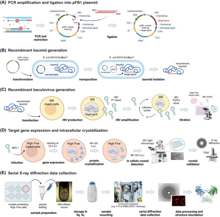
Crystallization of recombinant proteins in living cells is an emerging approach complementing conventional crystallization techniques. Homogeneous microcrystals well suited for serial diffraction experiments at X-ray free-electron lasers and synchrotron sources can be produced in a quasi-native environment, without the need for target protein purification. Several protein structures have already been solved; however, exploiting the full potential of this approach requires a systematic and versatile screening strategy for intracellular crystal growth. Recently, we published InCellCryst, a streamlined pipeline for producing microcrystals within living insect cells. Here, we present the detailed protocol, including optimized target gene expression using a baculovirus vector system, crystal formation, detection, and serial X-ray diffraction directly in the cells. The specific environment within the different cellular compartments acts as a screening parameter to maximize the probability of crystal growth. If successful, diffraction data can be collected 24 days after the start of target gene cloning.

Keywords: InCellCryst; X‐ray crystallography; baculovirus; in cellulo crystallization; protein crystallization; serial X‐ray diffraction.


Conflict of interest statement

The authors declare no conflict of interest.

Figures

Fig. 1
Fig. 1
Schematic summary of the InCellCryst pipeline. (A) The gene of interest is amplified by PCR and ligated into modified pFastBac1 plasmids. (B) After the transformation of E. coli DH10EmBacY cells, recombination with the bacmid takes place. The recombinant bacmid is isolated and (C) Sf9 insect cells are transfected for rBV generation. After high titer viral stock production, (D) High five insect cells are infected and used for high‐yield target gene expression. This eventually leads to the crystallization of the target protein within one of the cellular compartments, depending on the transport signal fused to the target protein sequence. (E) Crystal‐containing cells are directly used for serial diffraction data collection at 100 K at a synchrotron source. Serial diffraction data is finally processed using CrystFEL to elucidate the structure of the target protein. p.i., post‐infection; rBVs, recombinant baculoviruses. Created with BioRender.com.
Fig. 2
Fig. 2
Target gene cloning. (A) Schematic representation of modified pFB1 plasmids encoding cellular translocation sequences to transport the target protein co‐ or post‐translationally into cellular compartments that are characterized by individual environmental conditions. Cleavage with KpnI and NheI allows target gene cloning in frame with the start/stop codon and the translocation sequences encoded in the plasmids. Cleavage with BamHI and HindIII removes the entire cloning cassette from the plasmid. These pFB1 plasmids are also available encoding an additional HA tag (YPYDVPDYA) upstream of the KpnI site for N‐terminal fusion or downstream of the NheI site for C‐terminal fusion to the target protein. Moreover, a pFB1 cyto N‐His plasmid was designed to encode a His tag, an HA tag, and a TEV protease cleavage site upstream of the KpnI site. (B) Example of PCR primers for target gene amplification. At the 5′ ends, the fwd and rev primers need to encode a KpnI and NheI restriction site, respectively. Moreover, random nucleotides should be added at the ends to improve the efficiency of the PCR product cleavage. Created with BioRender.com. Full sequences of the presented pFB1 plasmids are provided as Supporting Information.
Fig. 3
Fig. 3
High Five insect cells containing ordered structures. Cells were infected with rBVs encoding (A) Trypanosoma brucei cathepsin B (CatB) [7, 23], (B) Trypanosoma brucei inosine monophosphate dehydrogenase (IMPDH) [18, 27], (C) Neurospora crassa HEX‐1 [15, 27], (D) Zika virus NS5 methyltransferase, (E) μNS protein from avian reovirus N‐terminally fused to GFP [29], and (F) Photinus pyralis luciferase [29] at an MOI of 1. Imaging followed 4 days post infection (dpi) i on a Nikon Ti2‐E or Ts2R‐FL microscope equipped with 100× objectives using the differential interference contrast (DIC) mode. Regular objects with sharp edges and mostly needle‐shaped morphology were detected (arrows). The translocation signal addressing a specific cellular compartment is indicated (cyto, cytosol; ER, endoplasmic reticulum; NLS, nucleus; PTS, peroxisomes). *, protein structure has already been elucidated using intracellularly grown crystals. Size bars for all images represent 20 μm.
Fig. 4
Fig. 4
Laboratory setup for MicroMesh loading. (A) A standard upright cell culture light microscope is equipped with a custom‐made holding device for a MicroMesh mounted on a magnetic goniometer base. A continuous humid air stream to keep the cells hydrated during loading is produced by passing compressed air through different water‐filled bottles (left). An empty bottle at the end prevents sample contamination with water droplets. (B) and (C) After adjusting the MeshMount in the optical focus of the microscope, the cells are directly pipetted onto the surface, followed by the removal of the excess liquid using an extra fine liquid wick from the back of the mesh. Immediately after applying cryo protection by pipetting 40% PEG200 on the loaded cells and blotting of exceeding liquids, the MeshMount is flash‐frozen in liquid N2.
Fig. 5
Fig. 5
Custom‐made coverslip‐holding device. (A) For crystal‐containing cell screening using an 100 × oil immersion objective and DIC, the glass coverslip with the adherent cells is transferred from the well of a 6‐well plate onto the lower metal ring structure (1). The upper plastic part is carefully screwed in (2) to seal the upper chamber. (B) The device is placed into a sample support, and 0.75 mL of ESF921 medium (+a/a) is pipetted into the chamber to keep the cells alive during light microscopy. (C) The assembled device is placed on the xy table of the Nikon Eclipse Ts2R microscope.

Similar articles

Cited by

References

    1. Schönherr R, Rudolph JM and Redecke L (2018) Protein crystallization in living cells. Biol Chem 399, 1–22. - PubMed
    1. Yamamoto M, Hirata K, Yamashita K, Hasegawa K, Ueno G, Ago H and Kumasaka T (2017) Protein microcrystallography using synchrotron radiation. IUCrJ 4, 529–539. - PMC - PubMed
    1. Yabashi M and Tanaka H (2017) The next ten years of X‐ray science. Nat Photon 11, 12–14.
    1. Standfuss J and Spence JCH (2017) Serial crystallography at synchrotrons and X‐ray lasers. IUCrJ 4, 100–101. - PMC - PubMed
    1. Spence JCH (2020) Serial crystallography: preface. Crystals 10, 135.

Substances

LinkOut - more resources