. 2025 Mar 28;21(3):e1012973.
doi: 10.1371/journal.ppat.1012973. eCollection 2025 Mar.

Dynamics of the adhesion complex of the human pathogens Mycoplasma pneumoniae and Mycoplasma genitalium

Affiliations

Dynamics of the adhesion complex of the human pathogens Mycoplasma pneumoniae and Mycoplasma genitalium

David Vizarraga et al. PLoS Pathog. .

Abstract

Mycoplasma pneumoniae and Mycoplasma genitalium are bacterial wall-less human pathogens and the causative agents of respiratory and reproductive tract infections. Infectivity, gliding motility and adhesion of these mycoplasmas to host cells are mediated by orthologous adhesin proteins forming a transmembrane adhesion complex that binds to sialylated oligosaccharides human cell ligands. Here we report the cryo-EM structure of M. pneumoniae P1 adhesin bound to the Fab fragment of monoclonal antibody P1/MCA4, which stops gliding and induces detachment of motile cells. The epitope of P1/MCA4 involves residues only from the small C-domain of P1. This epitope is accessible to antibodies only in the "closed conformation" of the adhesion complex and is not accessible in the "open" conformation, when the adhesion complex is ready for attachment to sialylated oligosaccharides. Polyclonal antibodies generated against the large N-domain of P1 or against the whole ectodomain of P40/P90 have little or no effects on adhesion or motility. Moreover, mutations in the highly conserved Engelman motifs found in the transmembrane helix of M. genitalium P110 adhesin also alter adhesion and motility. These results show that antibodies directed to the C-domain of P1 hinder the large conformational rearrangements in this domain required to alternate between the "open" and "closed" conformations of the adhesion complex. Since transition between both conformations is essential to complete the attachment/detachment cycle of the adhesion complex, interfering with the gliding of mycoplasma cells and providing a new potential target to confront M. pneumoniae and M. genitalium infections.


Conflict of interest statement

The authors have declared that no competing interests exist.

Figures
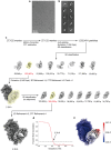
Fig 1
Fig 1. Cryo-EM structure determination of the P1-Fab(P1/MCA4) complex.
a) Representative micrograph of P1-Fab(P1/MCA4) complexes examined by CryoEM. b) Initial classes obtained after 2D classification of 2000 particles that were manually selected. c) Schematic processing overview for the P1-Fab(P1/MCA4) complex structure resolution from particles that were automatically picked from movies; CTF, Constrast transfer function. d) Fourier shell correlation of P1-Fab(P1/MCA4) complex at a final resolution of 2.39Å according to the 0.143 cut-off criteria. e) Final 3D reconstruction map color-coded according to local resolution.
Fig 2
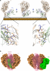
Fig 2. Cryo-EM structure of the P1-Fab(P1/MCA4) complex.
a) Ribbon representations, with three 90° apart views, of the complex between the ectodomain of P1 (brown) and the Fab fragment of Mab P1/MCA4 (light and heavy chains in blue and green, respectively). Insets indicate the main interactions between P1 and the Fab. The epitope is located in the C-domain of P1 closed to the mycoplasma membrane, which is also displayed as a reference. b) Two views of the Fab(P1/MCA4) (shown in green) and the extracellular region of the Nap, with P1 (brown) and P40/P90 (pink transparent). In the “open” conformation of the Nap (left panel) the P1/MCA4 epitope, buried at the center of the Nap, is totally inaccessible to antibodies and a Fab placed there would catastrophically clash with the adhesins. Therefore, to expose the P1/MCA4 epitope the C-domain of P1 has to experience an important rearrangement (right panel).
Fig 3
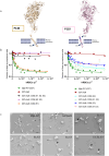
Fig 3. Engelman motifs in adhesins.
a) Transmembrane helices of adhesins from the M. pneumoniae cluster of mycoplasmas contain three highly conserved Engelman motifs (GxxxG sequences), referred as E1 (P140) and E2, E3 (P110) in M. genitalium. b) Mutational analysis, performed in the Engelman motifs of P140 and P110, indicate that motifs E2 and E3 from P110 have important and synergic contributions to adhesion. On the contrary, no effects were observed when E1 from P140 was mutated. c) Changes in adhesion correlate with increasing rates of cells presenting multiple TOs phenotypes.
Fig 4
Fig 4. The attachment and detachment cycle of the Nap complex.
An active Nap complex cycles between “open” and “closed” states, with the binding site to sialic oligosaccharides becoming alternatively accessible and inaccessible for binding. As shown in this work, during the nap cycle, structural rearrangements are experienced mainly by the C-domain of P1 (brown), with hinge rotations of about 175°, while the ectodomains of P40/P90 (pink) remain mostly unchanged. The wide movement of the C-domain from P1 drags, by contiguity, transmembrane helices thus reaching the internal parts of the TO (represented here simply as a line of elastic (red) and rigid (blue) components with possible displacements shown as black arrows). Binding of antibody P1/MCA (green) to the C-domain of P1 halts the cycle, trapping the “closed” conformation of the Nap. The structures known prior to this work are indicated using soft colors.

References

    1. Waites KB, Xiao L, Liu Y, Balish MF, Atkinson TP. Mycoplasma pneumoniae from the respiratory tract and beyond. Clin Microbiol Rev. 2017;30(3):747–809. doi: 10.1128/CMR.00114-16 - DOI - PMC - PubMed
    1. Parrott GL, Kinjo T, Fujita J. A compendium for Mycoplasma pneumoniae. Front Microbiol. 2016;7:513. doi: 10.3389/fmicb.2016.00513 - DOI - PMC - PubMed
    1. Rangroo R, Young M, Davis A, Pack S, Thakore S, Schepcoff A, et al.. the severity of the co-infection of Mycoplasma pneumoniae in COVID-19 patients. Cureus. 2022;14(4):e24563. doi: 10.7759/cureus.24563 - DOI - PMC - PubMed
    1. Meyer Sauteur PM, Beeton ML, European Society of Clinical Microbiology and Infectious Diseases (ESCMID) Study Group for Mycoplasma and Chlamydia Infections (ESGMAC), and the ESGMAC Mycoplasma pneumoniae Surveillance (MAPS) study group. Pneumonia outbreaks due to re-emergence of Mycoplasma pneumoniae. Lancet Microbe. 2024;5(6):e514. doi: 10.1016/S2666-5247(23)00406-8 - DOI - PubMed
    1. Mazzolini R, Rodríguez-Arce I, Fernández-Barat L, Piñero-Lambea C, Garrido V, Rebollada-Merino A, et al.. Engineered live bacteria suppress Pseudomonas aeruginosa infection in mouse lung and dissolve endotracheal-tube biofilms. Nat Biotechnol. 2023;41(8):1089–98. doi: 10.1038/s41587-022-01584-9 - DOI - PMC - PubMed

MeSH terms

Substances

LinkOut - more resources