. 2025 Mar 28;21(3):e1013014.
doi: 10.1371/journal.ppat.1013014. eCollection 2025 Mar.

RNAi screening of uncharacterized genes identifies promising druggable targets in Schistosoma japonicum

Affiliations

RNAi screening of uncharacterized genes identifies promising druggable targets in Schistosoma japonicum

Yuxiang Xie et al. PLoS Pathog. .

Abstract

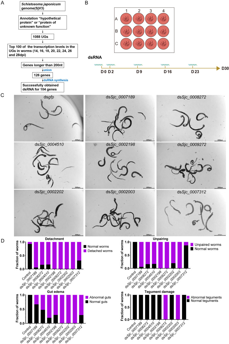
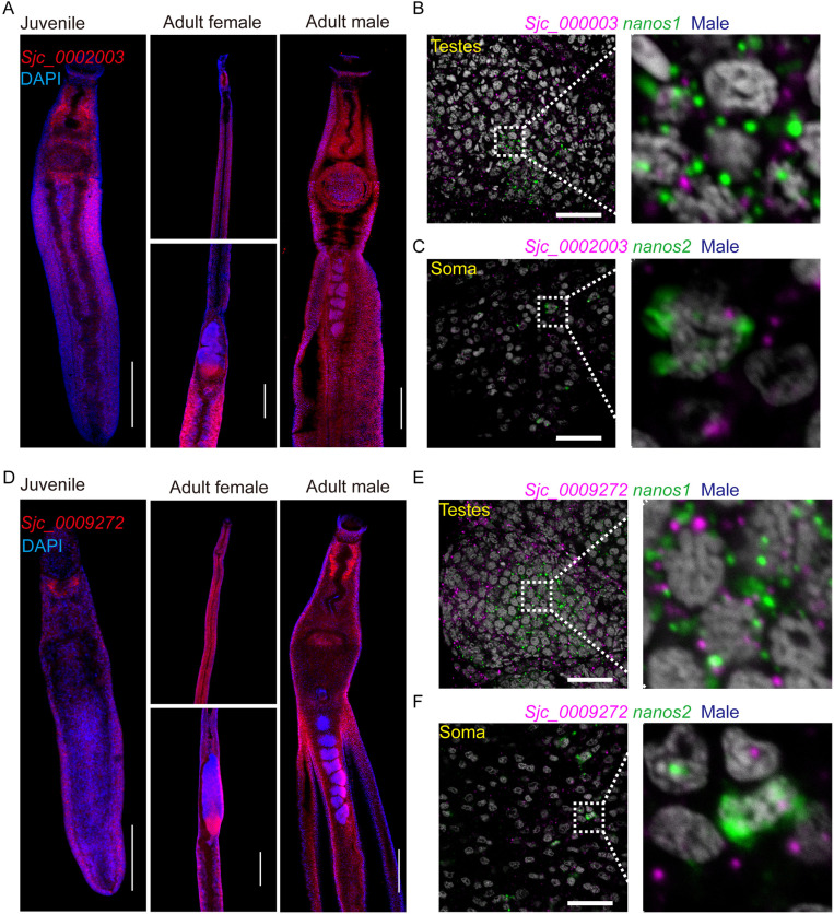
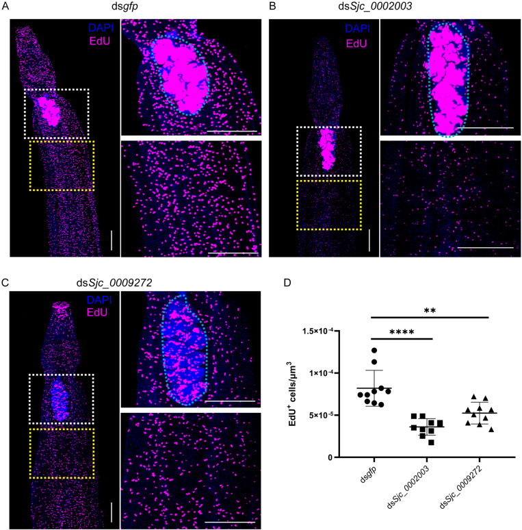
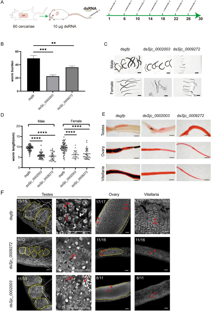
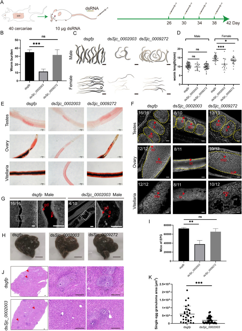
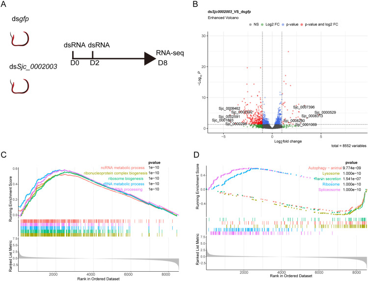
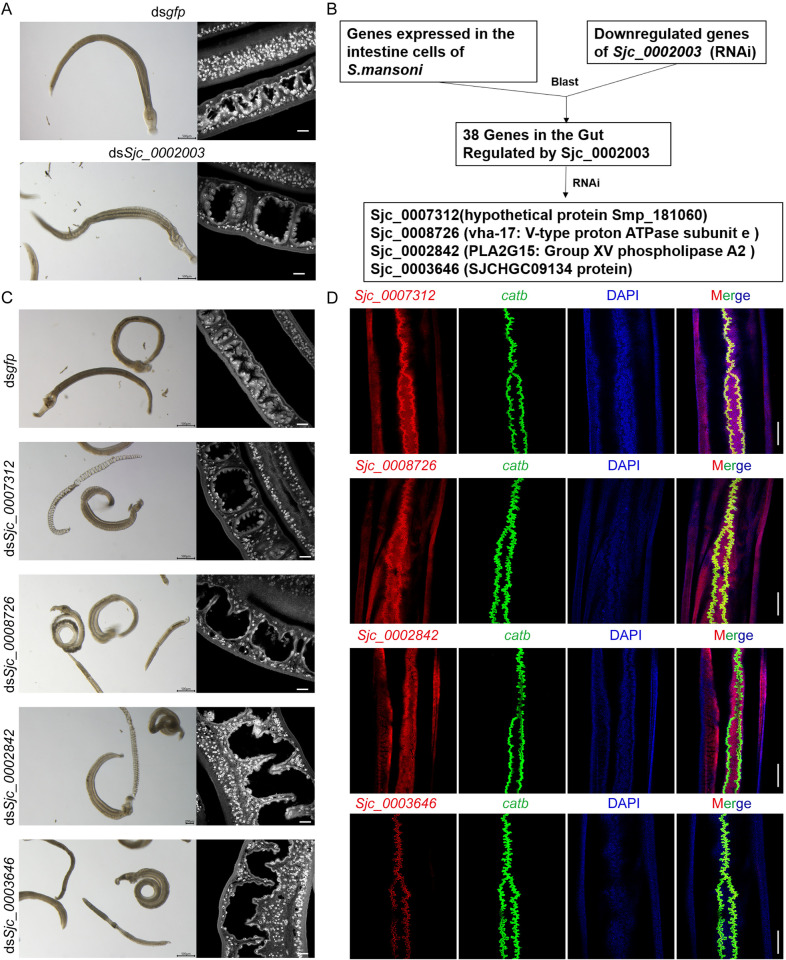
Schistosomiasis affects more than 250 million people worldwide and is one of the neglected tropical diseases. Currently, the treatment of schistosomiasis relies on a single drug-praziquantel-which has led to increasing pressure from drug resistance. Therefore, there is an urgent need to find new treatments. The development of genome sequencing has provided valuable information for understanding the biology of schistosomes. In the genome of Schistosoma japonicum, approximately 11% of the protein-coding sequences are uncharacterized genes (UGs) annotated as "hypothetical protein" or "protein of unknown function." These poorly understood genes have been unjustifiably neglected, although some may be essential for the survival of the parasites and serve as potential drug targets. In this study, we systematically mined the highly expressed UGs in both genders of this parasite throughout key developmental stages in their mammalian host, using our previously published S. japonicum genome and RNA-seq data. By employing in vitro RNA interference (RNAi), we screened 126 UGs that lack homologs in Homo sapiens and identified 8 that are essential for the parasite vitality. We further investigated two UGs, Sjc_0002003 and Sjc_0009272, which resulted in the most severe phenotypes. Fluorescence in situ hybridization demonstrated that both genes were expressed throughout the body without sex bias. Silencing either Sjc_0002003 or Sjc_0009272 reduced the cell proliferation in the body. Furthermore, in vivo RNAi indicated both genes are required for the growth and survival of the parasites in the mammalian host. For Sjc_0002003, we further characterize the underlying molecular cause of the observed phenotype. Through RNA-seq analysis and functional studies, we revealed that silencing Sjc_0002003 reduces the expression of a series of intestinal genes, including Sjc_0007312 (hypothetical protein), Sjc_0008276 (vha-17), Sjc_0002942 (PLA2G15), and Sjc_0003646 (SJCHGC09134 protein), leading to gut dilation. Our work highlights the importance of UGs in schistosomes as promising targets for drug development in the treatment of the schistosomiasis.


Conflict of interest statement

The authors have declared that no competing interests exist.

Figures

Fig 1
Fig 1. Eight UGs are essential for parasite vitality in vitro.
(A) Pipeline for the identification of target UGs. (B) Schematic diagram depicting the RNAi screening strategy in vitro. (C) Worms from the control group (dsgfp) and the eight UGs (RNAi) group at day 30 of in vitro culture, observed under light microscopy. Control (RNAi) worms remain paired and attached to the bottom of the plate. Worms in the eight UGs (RNAi) group show decreased activity and have lost the ability to attach. n ≥ 15 worm pairs, three biological replicates. Scale bars: 2000 μm. (D) Plots depicting the relative fraction of animals showing normal or defective phenotypes after following 30 days RNAi treatments. n ≥ 40 worm pairs, four biological replicates.
Fig 2
Fig 2. Localization of Sjc_0002003 and Sjc_0009272 mRNA in juvenile and mature worms.
(A) Fluorescence in situ hybridization showing mRNA expression of Sjc_0002003. Juvenile worms (left), adult female (middle) and adult male (right). For adult females, two areas are highlighted: the head (top) and the middle body including ovary and vitellaria (bottom). Juvenile worms, 14 dpi. Adult worms, 26-30 dpi. n ≥ 18, three biological replicates. Scale bars: 200 μm. (B-C) Double FISH of Sjc_0002003 with nanos1 (B)or nanos2 (C) in testes or soma of adult males. Right: magnified area in the white box on the left. Nuclei are shown in gray. n ≥ 8. Scale bars: 25 μm. (D) FISH showing mRNA expression of Sjc_0009272. n ≥ 18, three biological replicates. Scale bars: 200 μm. (E-F) Double FISH of Sjc_0009272 with nanos1 (E) or nanos2 (F) in testes or soma of adult males. Right: magnified area in the white box on the left. Nuclei are shown in gray. n ≥ 8. Scale bars: 25 μm.
Fig 3
Fig 3. Silencing Sjc_0002003 or Sjc_0009272 result in reduced cell proliferation.
(A-C) EdU labeling showing cell proliferation in control RNAi (A), Sjc_0002003 RNAi (B) and Sjc_0009272 RNAi (C) male parasites. Testes is in the white box. The blue dotted area is the testes. Yellow box indicates soma. Images are composed of multiple confocal stacks acquired from parasites 8 days after RNAi. Parasites were fixed after an overnight EdU labelling. Nuclei are shown in blue. EdU+ cells are in magenta. Scale bars: 200 μm. (D) Quantification of EdU+ cells per μm3 from the body in the yellow box. n = 10 worms. Each dot represents counts from a confocal stack taken from a single male parasite, with error bars representing 95% confidence intervals. Differences are statistically significant (****p < 0.0001, **p < 0.01, t-test).
Fig 4
Fig 4. Sjc_0002003 or Sjc_0009272 RNAi affects the growth, development, and survival of S. japonicum.
(A) Schematic diagram depicting the RNAi of schistosomula in vivo. On 1, 6, 10, 14, 18, 22, and 26 dpi, mice were injected in the tail vein with 10 μg of dsRNA targeting Sjc_0002003 or Sjc_0009272. gfp dsRNA was used as a negative control. On day 30, worms were harvested from the mice. (B) Worm burden of the parasites recovered at 30 dpi in control (RNAi), Sjc_0002003 (RNAi), and Sjc_0009272 (RNAi) groups. (C) Morphological observation of worms after RNAi. Scale bars: 2000 μm. (D) Worm length of the parasites recovered at 30 dpi in control (RNAi), Sjc_0002003 (RNAi), and Sjc_0009272 (RNAi) groups. (E) Carmine staining showing the reproductive organs under light microscopy. Scale bars: 200 μm. (F) Confocol microscopy of the testes, ovaries, and vitellaria in control (RNAi), Sjc_0002003 (RNAi), and Sjc_0009272 (RNAi) worms. so, spermatocyte; s, sperm; io, immature oocyte; mo, mature oocyte; iv, immature vitelline cell, mv, mature vitelline cell. Scale bars: 20 μm. Error bars represent 95% confidence intervals, n ≥ 3. Differences are statistically significant (****p < 0.0001, ***p < 0.001, **p < 0.01, t-test).
Fig 5
Fig 5. In vivo RNAi of Sjc_0002003 and Sjc_0009272 at the adult stage of S. japonicum.
(A) Schematic diagram depicting RNAi of adult parasites in vivo. On 26, 30, 34, and 38 dpi, mice were injected in the tail vein with 10 μg of dsRNA targeting the specified genes. At 42 dpi, worms were harvested. (B) Worm burden of the parasites recovered at 42 dpi in control (RNAi), Sjc_0002003 (RNAi), and Sjc_0009272 (RNAi) groups. 3 mice per group. (C) Morphological observation of worms after RNAi. Scale bars: 2000 μm. (D) Worm length of the parasites by sex recovered at 42 dpi. (E) Gonad observation in female and male worms by Carmine staining. Scale bars: 200 μm. (F) Carmine staining of the testes, ovaries, and vitellaria in worms from different RNAi treatment. so, spermatocyte; s, sperm; io, immature oocyte; mo, mature oocyte; iv, immature vitelline cell; mv, mature vitelline cell. Scale bars: 20 μm. (G) Confocal imaging of the parasite gut in Carmine-stained males following Sjc_0002003 silencing. g, gut; gd, gastrodermis. Scale bars: 25 μm. (H) Gross observations of the mouse liver from the dsgfp and dsSjc_0002003 treatment groups. Scale bars: 1 cm. (I) Egg count per gram of liver after RNAi. 3 livers per group. (J) Histological assessment of mouse liver by H&E (hematoxylin and eosin) staining. Scale bars: 200 μm. (K) Statistical analysis of the size of egg granuloma area after RNAi. 3 liver sections per group. Error bars represent 95% confidence intervals, n ≥ 3. ‘ns’ indicates no significant difference (p > 0.05). Differences are statistically significant (***p < 0.001, **p < 0.01, *p < 0.05, t-test).
Fig 6
Fig 6. GO and KEGG pathway analysis results from GSEA.
(A) RNAi strategy for RNA-seq: After 8 days of RNAi, paired worms were separated, and male parasites were harvested for RNA-seq. (B) Volcano plot illustrating differentially expressed genes (DEGs) in male parasites. DEGs were defined as |Log2 Fold Change| ≥1 and adjusted P-value< 0.05. (C) GSEA enrichment analysis of the expression data in males, focusing on Biological Processes. (D) GSEA enrichment analysis of the expression data in males, highlighting KEGG Pathway Analysis. Gene sets were considered significant only when |NES| > 1. The top 5 leading gene sets are displayed in the plot. GSEA, Gene Set Enrichment Analysis; GO, Gene Ontology; NES, normalized enrichment score.
Fig 7
Fig 7. Sjc_0002003 modulates intestinal function through the regulation of intestinal genes.
(A) Images of worms under light microscopy (left) and confocal images (right) of the Carmine-stained males after 12 days of RNAi treatment in vitro. Scale bars: 500 μm (left); 20 μm (right). (B) Schematic representation of the functional analysis of DEGs downregulated by Sjc_0002003 RNAi. (C) Images of worms under light microscopy (left) and confocal images (right) of the Carmine-stained males after 30 days of RNAi treatment in vitro. Scale bars: 500 μm (left); 20 μm (right). (D) Colocalization of Sjc_0007312, Sjc_0008726, Sjc_0002842 or Sjc_0003646 with the intestinal marker catb. Scale bars: 200 μm.

Similar articles

References

    1. Mutapi F, Maizels R, Fenwick A, Woolhouse M. Human schistosomiasis in the post mass drug administration era. Lancet Infect Dis. 2017;17(2):e42–8. doi: 10.1016/S1473-3099(16)30475-3 - DOI - PMC - PubMed
    1. Malta KK, Silva TP, Palazzi C, Neves VH, Carmo LAS, Cardoso SJ, et al.. Changing our view of the Schistosoma granuloma to an ecological standpoint. Biol Rev Camb Philos Soc. 2021;96(4):1404–20. doi: 10.1111/brv.12708 - DOI - PubMed
    1. Ismail M, Botros S, Metwally A, William S, Farghally A, Tao LF, et al.. Resistance to praziquantel: direct evidence from Schistosoma mansoni isolated from Egyptian villagers. Am J Trop Med Hyg. 1999;60(6):932–5. doi: 10.4269/ajtmh.1999.60.932 - DOI - PubMed
    1. Wang J, Paz C, Padalino G, Coghlan A, Lu Z, Gradinaru I, et al.. Large-scale RNAi screening uncovers therapeutic targets in the parasite Schistosoma mansoni. Science. 2020;369(6511):1649–53. doi: 10.1126/science.abb7699 - DOI - PMC - PubMed
    1. Adhikari S, Nice EC, Deutsch EW, Lane L, Omenn GS, Pennington SR, et al.. A high-stringency blueprint of the human proteome. Nat Commun. 2020;11(1):5301. doi: 10.1038/s41467-020-19045-9 - DOI - PMC - PubMed

Substances

LinkOut - more resources