Observational Study
. 2025 Jul;211(7):1253-1263.
doi: 10.1164/rccm.202409-1792OC.

Dynamic 11C- Para-Aminobenzoic Acid Positron Emission Tomography/Computed Tomography for Visualizing Pulmonary Mycobacteroides abscessus Infections

Affiliations
Observational Study

Dynamic 11C- Para-Aminobenzoic Acid Positron Emission Tomography/Computed Tomography for Visualizing Pulmonary Mycobacteroides abscessus Infections

Yuderleys Masias-Leon et al. Am J Respir Crit Care Med. 2025 Jul.

Abstract

Rationale: Mycobacteroides abscessus infections affect immunocompromised patients and those with underlying pulmonary disease. Conventional imaging cannot distinguish M. abscessus infections from underlying pulmonary disease or sterile inflammation, requiring invasive procedures for definitive diagnosis. Objectives: We evaluated 11C-para-aminobenzoic acid (11C-PABA), a chemically identical radioanalog of PABA, to detect and localize infections due to M. abscessus. Methods: In vitro uptake assays were performed to test the metabolism and accumulation of PABA into M. abscessus reference and clinical isolates. Dynamic 11C-PABA positron emission tomography (PET) was performed in a mouse model of M. abscessus pulmonary infection and in a patient with microbiologically confirmed M. abscessus pulmonary infection (NCT05611905). Measurements and Main Results: 11C-PABA was intracellularly metabolized by M. abscessus to 11C-7,8-dihydropteroate. In addition, the reference strain and all 13 randomly chosen clinical isolates, including 3 resistant to trimethoprim-sulfamethoxazole, rapidly accumulated PABA. No PABA accumulation was noted by heat-inactivated bacteria or mammalian cells. Dynamic 11C-PABA PET in a mouse model of M. abscessus pulmonary infection rapidly distinguished infection from sterile inflammation and also accurately monitored response to antibiotic treatment. Finally, dynamic 11C-PABA PET in a 33-year-old woman with cystic fibrosis and microbiologically confirmed M. abscessus pulmonary infection was safe and demonstrated significantly higher and sustained PET uptake in the affected lesions. Conclusions: 11C-PABA PET is an innovative, clinically translatable, noninvasive, bacteria-specific diagnostic to differentiate M. abscessus infections from underlying pulmonary disease in patients. This tool could also help in monitoring treatment responses and enable precision medicine approaches for patients with complicated infections.

Keywords: molecular imaging; nontuberculous mycobacteria; pathogen specific.


Figures
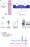
Figure 1.
Figure 1.
11C–para–aminobenzoic acid (11C-PABA) metabolism in Mycobacteroides abscessus. (A) Schematic representation of the intrabacterial incorporation of 11C-PABA (chemically identical to PABA) by dihydropteroate synthase (DHPS) via the use of DHPPP into 11C-dihydropteroate (11C-DHP). (B) Methodology for evaluating PABA metabolism in M. abscessus. (C) Radio-HPLC studies of lysed bacterial pellets demonstrate conversion of 11C-PABA into 11C-DHP, which is the final product of the enzymatic reaction catalyzed by the bacterial DHPS. The x-axis indicates the retention time in minutes. At least three radio-HPLC runs were performed for each compound. DHPPP = 6-hydroxymethyl-7,8-dihydropterin-pyrophosphate.
Figure 2.
Figure 2.
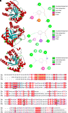
Docking model. (A) Proposed interactions from the docking of PABA with Bacillus anthracis (Ba) (PDB: 3TYB). (B) Proposed interactions from the docking of PABA with DHPS from Escherichia coli (Ec) (PDB: 1AJ0). (C) Proposed interactions from the docking of PABA with DHPS from Mycobacteroides abscessus (Ma) (SWISS-MODEL). (D) Alignment of the FASTA sequence from Ba, Ec, and Ma DHPS and their respective binding amino acids (arrowheads). Highlighted colors represent the amino acid residues predicted to form loop 1 (yellow), loop 2 (blue) and α-loop7 (pink). Red letters and blue frames represent similarity within and across groups, respectively. DHPS = dihydropteroate synthase; PABA = para–aminobenzoic acid; PDB = Protein Data Bank.
Figure 3.
Figure 3.
Para–aminobenzoic acid (PABA) uptake by Mycobacteroides abscessus. (A) 3H-PABA uptake after six hours of incubation with live (blue) and heat-inactivated (red) M. abscessus (reference, ATCC 19977) cultures. (B) Competitive inhibition of 3H-PABA uptake with increasing concentrations of unlabeled PABA. (C) Antimicrobial resistance pattern for the T/S-resistant M. abscessus clinical isolates (strain IDs 362 and 361 are subspecies massilliense, and 972 is subspecies abscessus). (D) 3H-PABA uptake by the reference and clinical isolates. No uptake was noted in the heat-inactivated cultures (red) or mammalian cells (ATCC J774.1, green). Data are expressed as median and interquartile range. AMI = amikacin; ATCC = American Type Culture Collection; CFX = cefoxitin; CIP = ciprofloxacin; CLR = clarithromycin; I = intermediate; ID = identifier; IMI = imipenem; LZD = linezolid; MDR = multidrug resistant; MXF = moxifloxacin; NR = not run; R = resistant; S = susceptible; TOB = tobramycin; T/S = trimethoprim/sulfamethoxazole.
Figure 4.
Figure 4.
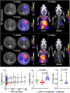
PET/CT in mice. (A–C) Representative transverse sections from dynamic 11C–para–aminobenzoic acid (11C-PABA) PET/CT (A), 18F–fluorodeoxyglucose (FDG)–PET/CT (B), and maximum-intensity projections showing the first (0–10 min) and last (50–60 min) frames from the dynamic 11C-PABA PET (C) in mice with Mycobacteroides abscessus pulmonary infection are shown. (D–F) Representative transverse sections from dynamic 11C-PABA PET/CT (D), 18F-FDG-PET/CT (E), and maximum-intensity projections showing the first (0–10 min) and last (50–60 min) frames from the dynamic 11C-PABA PET (F) in mice with LPS-induced sterile pulmonary inflammation are shown. Pulmonary lesions are marked with arrows. (G and H) Pulmonary 11C-PABA PET time–activity curves normalized to blood (G), and AUCr (H) are shown (n = 7 animals for the M. abscessus pulmonary infection group and n = 4 animals for the LPS-induced sterile pulmonary inflammation group; affected and unaffected lung areas are from mice with M. abscessus pulmonary infection; two VOIs per animal). (I) Pulmonary 18F-FDG-PET SUV is shown (n = 4 animals each for the M. abscessus pulmonary infection group and LPS-induced sterile pulmonary inflammation groups; two VOIs per animal). Data are expressed as median and interquartile range. Statistical analyses were performed using a two-tailed Mann-Whitney U test. AUCr = area under the curve ratio of lesion to blood; CT = computed tomography; H = heart; PET = positron emission tomography; SUV = mean standardized uptake value; SUVr = mean standardized uptake value ratio; VOI = volume of interest.
Figure 5.
Figure 5.
Monitoring response to antibiotic treatments using 11C–para–aminobenzoic acid (11C-PABA) positron emission tomography (PET)/computed tomography (CT). (A and B) Representative 11C-PABA PET/CT transverse (top) and coronal (bottom) images from mice with Mycobacteroides abscessus pulmonary infection (A) and those with antibiotic (clofazimine) treatment for 4 weeks (B) are shown. (C and D) Pulmonary 11C-PABA PET time–activity curves normalized to blood (C), and the AUCr (D) are shown (n = 7 animals for the M. abscessus pulmonary infection group and n = 4 animals for the antibiotic treated group; two VOIs per animal). Pulmonary lesions are marked with arrows. Data are expressed as median and interquartile range. Statistical analyses were performed using a two-tailed Mann-Whitney U test. AUCr = area under the curve ratio of lesion to blood; H = heart; SUVr = mean standardized uptake value ratio; VOI = volume of interest.
Figure 6.
Figure 6.
Dynamic 11C-PABA positron emission tomography (PET) in a patient with pulmonary Mycobacteroides abscessus infection. (A and B) Representative coronal (left) and transverse (right) computed tomography (CT) and 11C-PABA PET/CT images (A) and 11C-PABA PET area under the curve (AUC) heatmaps (B) from a 33-year-old woman with cystic fibrosis and microbiologically confirmed M. abscessus infection. Pulmonary lesions are marked with arrows, and the numbers in A indicate minutes after tracer injection. (C and D) 11C-PABA PET time–activity curves (C) and AUC (D) over 45 minutes in the affected and unaffected lung regions. Data are expressed as median and interquartile range. Statistical analyses were performed using a two-tailed Mann-Whitney U test. 11C-PABA = 11C–para–aminobenzoic acid; H = heart; L = liver; V = vessels.

Comment in

References

    1. Baldwin SL, Larsen SE, Ordway D, Cassell G, Coler RN. The complexities and challenges of preventing and treating nontuberculous mycobacterial diseases. PLoS Negl Trop Dis . 2019;13:e0007083. - PMC - PubMed
    1. Wu ML, Aziz DB, Dartois V, Dick T. NTM drug discovery: status, gaps and the way forward. Drug Discov Today . 2018;23:1502–1519. - PMC - PubMed
    1. Winthrop KL, Marras TK, Adjemian J, Zhang H, Wang P, Zhang Q. Incidence and prevalence of nontuberculous mycobacterial lung disease in a large US managed care health plan, 2008–2015. Ann Am Thorac Soc . 2020;17:178–185. - PMC - PubMed
    1. Falkinham JO., III Environmental sources of nontuberculous mycobacteria. Clin Chest Med . 2015;36:35–41. - PubMed
    1. Johansen MD, Herrmann JL, Kremer L. Non-tuberculous mycobacteria and the rise of Mycobacterium abscessus. Nat Rev Microbiol . 2020;18:392–407. - PubMed

Publication types

MeSH terms