. 2025 Mar 28;16(1):3047.
doi: 10.1038/s41467-025-58264-w.

Structural visualization of small molecule recognition by CXCR3 uncovers dual-agonism in the CXCR3-CXCR7 system

Affiliations

Structural visualization of small molecule recognition by CXCR3 uncovers dual-agonism in the CXCR3-CXCR7 system

Shirsha Saha et al. Nat Commun. .

Abstract

Chemokine receptors are critically involved in multiple physiological and pathophysiological processes related to immune response mechanisms. Most chemokine receptors are prototypical GPCRs although some also exhibit naturally-encoded signaling-bias toward β-arrestins (βarrs). C-X-C type chemokine receptors, namely CXCR3 and CXCR7, constitute a pair wherein the former is a prototypical GPCR while the latter exhibits selective coupling to βarrs despite sharing a common natural agonist: CXCL11. Moreover, CXCR3 and CXCR7 also recognize small molecule agonists suggesting a modular orthosteric ligand binding pocket. Here, we determine cryo-EM structures of CXCR3 in an Apo-state and in complex with small molecule agonists biased toward G-proteins or βarrs. These structural snapshots uncover an allosteric network bridging the ligand-binding pocket to intracellular side, driving the transducer-coupling bias at this receptor. Furthermore, structural topology of the orthosteric binding pocket also allows us to discover and validate that selected small molecule agonists of CXCR3 display robust agonism at CXCR7. Collectively, our study offers molecular insights into signaling-bias and dual agonism in the CXCR3-CXCR7 system with therapeutic implications.


Conflict of interest statement

Competing interests: The authors declare no competing interests.

Figures
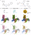
Fig. 1
Fig. 1. Overall architecture of VUF11418-bound, VUF10661-bound, and Apo-CXCR3-G-protein complexes.
a, b Chemical structure of VUF11418 (a) and VUF10661 (b). Chemical structures have been prepared using Marvin JS. ce Map and ribbon diagram of the ligand-bound and Apo-CXCR3-Go complexes (front view) and the cryo-EM densities of the ligands (sticks) are depicted as transparent surface representations. (VUF11418-CXCR3: pale violet red, VUF11418: deep teal, VUF10661-CXCR3: olive drab, VUF10661: cyan, Apo-CXCR3: dark goldenrod, miniGαo: cornflower blue, Gβ1: light coral, Gɣ2: chartreuse, scFv16: gray).
Fig. 2
Fig. 2. Ligand binding interface and aromatic cage in CXCR3.
a, b Cross-section of the orthosteric pocket in CXCR3 depicting the aromatic cage and the depth of penetration of the ligands with respect to the conserved W6.48. c Orthosteric pocket in CXCR3 showing bound VUF11418 and VUF10661 in inverted ‘U’ and linear shaped conformations, respectively. d, e List of CXCR3 residues interacting with the ligands VUF11418 and VUF10661. f Rotameric transitions in Apo-CXCR3 when compared with the inactive state and VUF-bound CXCR3 structures. (AMG487-CXCR3: purple; PDB: 8K2W, VUF11418-CXCR3: pale violet red, VUF11418: deep teal, VUF10661-CXCR3: olive drab, VUF10661: cyan, Apo-CXCR3: dark goldenrod).
Fig. 3
Fig. 3. Effect of mutating CXCR3 ligand interacting residues on transducer coupling.
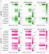
a, b Heatmap showing G-protein signaling, as measured using cAMP secondary messenger response (Gi-mediated decrease in cytosolic cAMP levels) and GoB dissociation (indicated by a decrease in luminescence), and βarr1 recruitment (indicated by an increase in luminescence) downstream to CXCR3 mutants following stimulation with VUF10661 (green) and VUF11418 (pink). Data (mean ± SEM) represents either three (for G-protein assays) or four (for βarr1 recruitment) independent experiments, performed in duplicate, and has been normalized with respect to the signal observed in unstimulated condition, treated as either 100% (for G-protein assays) or 1 (for βarr1 recruitment). Source data are provided as a Source Data file.
Fig. 4
Fig. 4. W1092.60 exerts a distinct effect in regulating signaling downstream to both VUFs.
a Stimulation of CXCR3W109A (red) with VUF10661 fails to elicit any response in G-protein signaling and βarr1 recruitment assays, while VUF11418 induces a slightly enhanced response, as compared to CXCR3WT (black). Data (mean ± SEM) represents three (for G-protein assays) or five (for βarr1 recruitment) independent experiments, performed in duplicate, and has been normalized with respect to the signal observed at the lowest ligand concentration, treated as either 100% (for G-protein assays) or 1 (for βarr1 recruitment). b W1092.60 in CXCR3 interacts with VUF10661, while rotation of this residue in the VUF11418-bound structure prevents its interaction with VUF11418. (VUF10661-CXCR3: olive drab, VUF10661: cyan, VUF11418-CXCR3: pale violet red, VUF11418: deep teal). Source data are provided as a Source Data file.
Fig. 5
Fig. 5. Mutating Y2716.51 leads to differential loss of signaling.
a Stimulation of CXCR3Y271A (blue) with either VUF10661 or VUF11418 induces G-protein signaling to a similar extent as wild-type receptor (black) when measured using a cAMP response assay, and reduced G-protein dissociation. Stimulation of CXCR3Y271A with VUF10661 and VUF11418 leads to a drastic loss in βarr1 recruitment. Data (mean ± SEM) represents either three (for G-protein assays) or four (for βarr1 recruitment) independent experiments, performed in duplicate, and has been normalized with respect to the signal observed at the lowest ligand concentration, treated as either 100% (for G-protein assays) or 1 (for βarr1 recruitment). b Y2716.51 adopts a similar orientation and contacts the ligand in both VUF11418 and VUF10661-bound CXCR3 structures. (VUF10661-CXCR3: olive drab, VUF10661: cyan, VUF11418-CXCR3: pale violet red, VUF11418: deep teal). Source data are provided as a Source Data file.
Fig. 6
Fig. 6. Comparative analysis of CXCR3 structures.
a Bottom view (left), side view (middle), and top view (right) of CXCR3 depicting structural reorientation upon binding VUF11418 (deep teal) and VUF11222 (violet). b Flip observed in VUF11418 and VUF11222 to contact nearby CXCR3 residues. c Rotational shift in W1092.60 to avoid a steric clash with the small molecule agonists. d Rotational shift in the side chains of the residues constituting the aromatic cage around the ligand. e Shift of ICL1 away from the cytoplasmic core in VUF11418-bound CXCR3. f Opening up of helical conformation in ICL2 of VUF11418-bound CXCR3 and movement toward the cytoplasmic core. g Structural superimposition of VUF10661-CXCR3 and PS372424-CXCR3. hk Major interacting residues in CXCR3 contacting VUF10661 (cyan) and PS372424 (coral). l, m Shift in ICL2 (l) and ICL3 (m) of VUF10661-bound CXCR3 as compared to PS372424-bound CXCR3. n Structural superimposition of Apo-CXCR3 and CXCL10-bound CXCR3. o, p Transmembrane helices move away from the orthosteric pocket in the Apo-CXCR3 structure. qt Side chain rearrangements in the key residues as observed in Apo-CXCR3 and CXCL10-CXCR3. (VUF11418-CXCR3: pale violet red, VUF11222-CXCR3: light beige; PDB: 8HNM, VUF10661-CXCR3: olive drab, PS372424-CXCR3: pale violet; PDB: 8HNL, Apo-CXCR3: dark goldenrod, CXCL10-CXCR3: sky blue; PDB: 8K2X, CXCL10: chartreuse).
Fig. 7
Fig. 7. Major conformational changes on CXCR3 activation.
a Superimposition of inactive CXCR3 with Apo-state receptor and receptor bound to VUF11418 and VUF10661. b, c Displacements of TM1, TM2, TM3, TM6, TM7 and helix8 upon CXCR3 activation in the structures of Apo-CXCR3, VUF11418-CXCR3, VUF10661-CXCR3, respectively. d Conformational changes in the conserved microswitches (DRY, PA(I)F, NPxxY, CWxP) in the structures of CXCR3. (Apo-CXCR3: dark goldenrod, VUF11418-CXCR3: pale violet red, VUF10661-CXCR3: olive drab, AMG487-CXCR3: purple; PDB: 8K2W).
Fig. 8
Fig. 8. Allosteric network in VUF10661-bound CXCR3.
a Residues promoting allosteric communication in VUF10661-CXCR3 exhibit different orientations than those in VUF11418-CXCR3. Superimposition of the two VUF-bound structures reveals an RMSD of ~1.2 Å. b Key residues in CXCR3 promoting allosteric communication. c Allosteric signal propagation in VUF10661-CXCR3. (VUF11418-CXCR3: pale violet red, VUF10661-CXCR3: olive drab).
Fig. 9
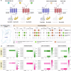
Fig. 9. VUF11418 and VUF10661 activate signaling downstream to CXCR7.
a Schematic representation of canonical signaling downstream of CXCR3 and CXCR7 stimulation with chemokines and synthetic agonists. Created in BioRender. #3, G. (2025) https://BioRender.com/r69j488. b Cross-section of the ligand binding pocket in CCX662 bound CXCR7 (PDB: 7SK9). c Conserved interacting sites in VUF11418-CXCR3, VUF10661-CXCR3 and CCX662-CXCR7. d Heatmap showing VUF10661 (green) and VUF11418 (pink) selectivity across all CXCRs in inducing cAMP signaling (Gi-mediated decrease in cytosolic cAMP), GoB dissociation (as measured by a decrease in luminescence) and βarr1/2 recruitment (as measured by an increase in luminescence). Data (mean) represents three independent biological replicates, performed in duplicate, and normalized with respect to signal observed in the absence of stimulation, treated either as 100% (for cAMP response), or 1 (for GoB dissociation and βarr1/2 recruitment). Source data are provided as a Source Data file.
Fig. 10
Fig. 10. Validation of dual-agonism of VUFs at CXCR7.
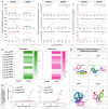
a VUF10661 (green) and VUF11418 (pink) stimulate both CXCR3 and CXCR7 as measured in various assays. Data (mean ± SEM) represents either three (for βarr1/2 recruitment) or four (for cAMP response assay) independent biological replicates, performed in duplicate, and normalized with respect to signal observed at the lowest dose, treated either as 100% (for cAMP response), or 1 (βarr1/2 recruitment). VUF11207 (orange) has been previously characterized to be specific for CXCR7. b Heatmap showing βarr1 recruitment downstream to CXCR7 mutants following stimulation with VUF10661 (green) and VUF11418 (pink). Data (mean ± SEM) represents three independent experiments, performed in duplicate, and has been normalized with respect to the signal observed under unstimulated condition, treated as 1. c Mutating Y268A (blue) leads to a drastic reduction in βarr1 recruitment following stimulation with both VUFs, whereas mutating L128A (orange) ablates βarr1 recruitment upon stimulation with VUF10661 while eliciting only marginally reduced βarr1 recruitment following VUF11418 stimulation, as compared to wild-type CXCR7 (purple). Data (mean ± SEM) represents four independent experiments, performed in duplicate, and has been normalized with respect to the signal observed under unstimulated conditions, treated as 1. d Residues promoting allosteric communication in VUF10661-CXCR3 (green) exhibit similar rotameric shifts with respect to CCX662-CXCR7 (blue, PDB: 7SK9) and different orientations than those in VUF11418-CXCR3 (pink). Source data are provided as a Source Data file.

References

    1. Hughes, C. E. & Nibbs, R. J. B. A guide to chemokines and their receptors. FEBS J.285, 2944–2971 (2018). - PMC - PubMed
    1. Viola, A. & Luster, A. D. Chemokines and their receptors: drug targets in immunity and inflammation. Annu Rev. Pharmacol. Toxicol.48, 171–197 (2008). - PubMed
    1. Scholten, D. J. et al. Pharmacological modulation of chemokine receptor function. Br. J. Pharmacol.165, 1617–1643 (2012). - PMC - PubMed
    1. Rossi, D. & Zlotnik, A. The biology of chemokines and their receptors. Annu Rev. Immunol.18, 217–242 (2000). - PubMed
    1. O’Hayre, M., Salanga, C. L., Handel, T. M. & Allen, S. J. Chemokines and cancer: migration, intracellular signalling and intercellular communication in the microenvironment. Biochem J.409, 635–649 (2008). - PubMed

LinkOut - more resources