

The value of predicting breast cancer with a DBT 2.5D deep learning model
- PMID: 40155449
- PMCID: PMC11953484
- DOI: 10.1007/s12672-025-02170-6
The value of predicting breast cancer with a DBT 2.5D deep learning model
Abstract
Objective: To evaluate the accuracy and efficacy of a 2.5-dimensional deep learning (DL) model based on digital breast tomosynthesis (DBT) in predicting breast cancer.
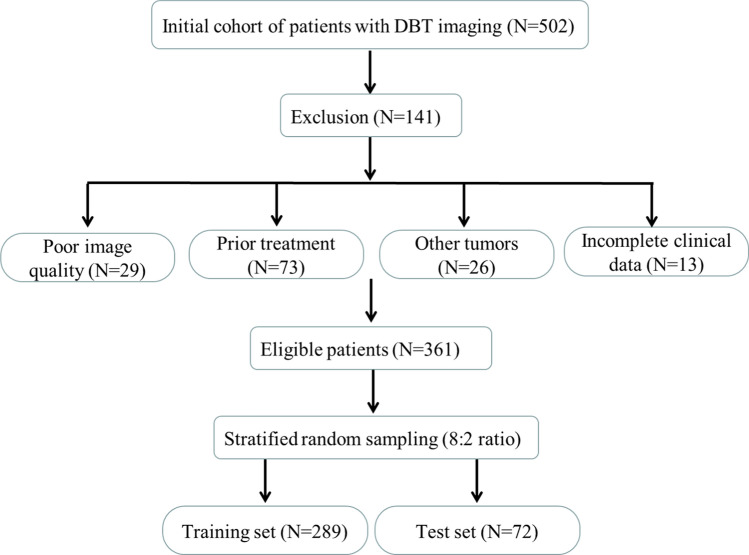
Methods: Through a retrospective analysis of data from 361 patients with breast tumor lesions treated at Shandong Provincial Hospital Affiliated to Shandong First Medical University between 2018 and 2020, this study utilized deep convolutional neural networks (DCNN) to automatically extract key features from DBT images. By applying dimensionality reduction and feature fusion selection, a variety of machine learning predictive models based on a 2.5-dimensional feature set were constructed. Additionally, a comprehensive predictive model was developed by combining univariate and multivariate logistic regression analyses with clinical data. The model's performance was assessed using receiver operating characteristic (ROC) curves, area under the curve (AUC) values, and accuracy rates.
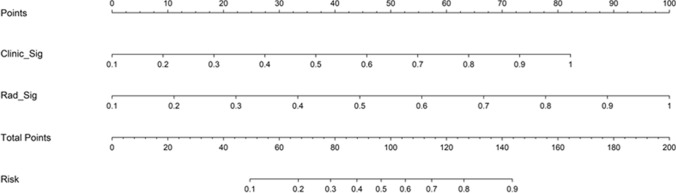
Results: In the test set, DBT 2.5D deep learning-based logistic regression, LightGBM, multilayer perceptron, and comprehensive models achieved accuracies of 72.2%, 75.0%, 79.2%, and 80.6%; AUCs of 0.826, 0.756, 0.859, and 0.871; sensitivities of 63.8%, 70.2%, 80.9%, and 87.2%; specificities of 88.0%, 84.0%, 76.0%, and 68.0%; PPVs of 90.9%, 89.2%, 86.4%, and 83.7%; NPVs of 56.4%, 60.0%, 67.9%, and 73.9%; and F1 scores of 75.0%, 78.6%, 83.5%, and 85.4%, respectively. These results underscore the high efficiency and potential of DBT 2.5D deep learning models in breast cancer diagnosis, particularly the comprehensive model's superior performance across key metrics.
Conclusion: The 2.5D deep learning model based on DBT shows good performance in preoperative breast cancer prediction, with its integration with clinical data further enhancing its effectiveness. The combination of deep learning and radiomics offers a viable approach for early breast cancer diagnosis, supporting the development of more accurate personalized diagnostic and treatment strategies.
Keywords: 2.5D deep learning; Breast cancer; Diagnosis; Digital breast tomosynthesis; Predictive model.
© 2025. The Author(s).
Conflict of interest statement
Declarations. Ethics approval and consent to participate: Ethical review and approval were waived for this study due to the Ethics Committee of Shandong Provincial Hospital. This study was conducted in accordance with the principles of the Declaration of Helsinki. Informed consent: Informed consent was obtained from all subjects involved in the study. Written informed consent has been obtained from the patients to publish this paper. Competing interests: The authors declare no competing interests.
Figures


References
-
- Sung H, Ferlay J, Siegel RL, et al. Global Cancer Statistics 2020: GLOBOCAN estimates of incidence and mortality worldwide for 36 cancers in 185 countries. CA Cancer J Clin. 2021;71(3):209–49. - PubMed
-
- Preibsch H, Siegmann-Luz KC. Digitale Tomosynthese der Mamma [Digital breast tomosynthesis]. Radiologe. 2015;55(1):59–67 (quiz 68-70). - PubMed
-
- Slanetz PJ. Digital breast tomosynthesis screening for breast cancer: it is cost-effective! Radiology. 2020;297(1):49–50. - PubMed
-
- Currie G, Hawk KE, Rohren E, et al. Machine learning and deep learning in medical imaging: intelligent imaging. J Med Imaging Radiat Sci. 2019;50(4):477–87. - PubMed
LinkOut - more resources
Full Text Sources