. 2025 Mar 28;11(1):61.
doi: 10.1038/s41531-025-00905-4.

The homozygous LRRK2.p.N1437D point mutation mouse is a novel model of parkinsonism

Affiliations

The homozygous LRRK2.p.N1437D point mutation mouse is a novel model of parkinsonism

Lin-Hua Gan et al. NPJ Parkinsons Dis. .

Abstract

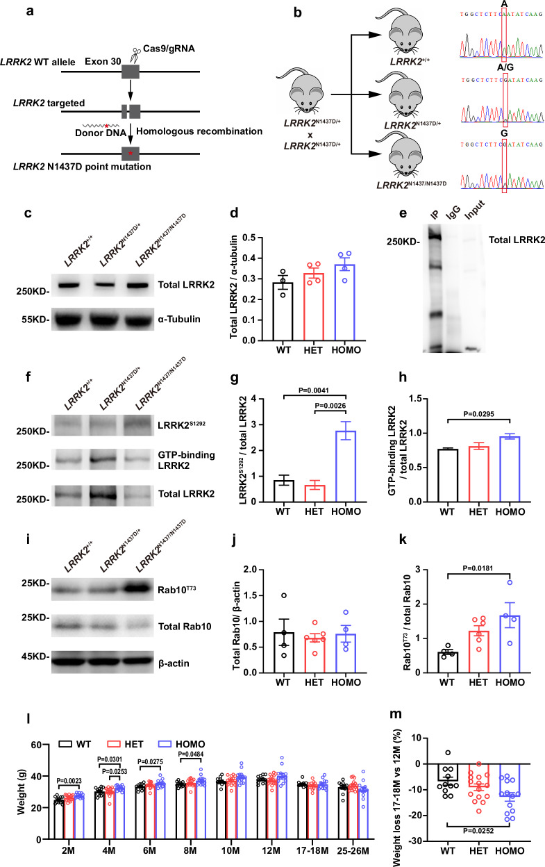
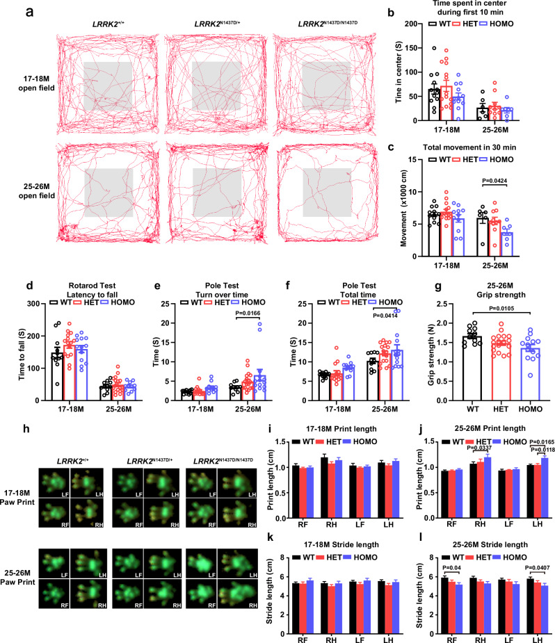
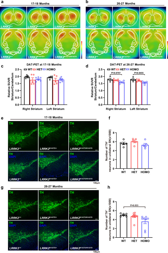
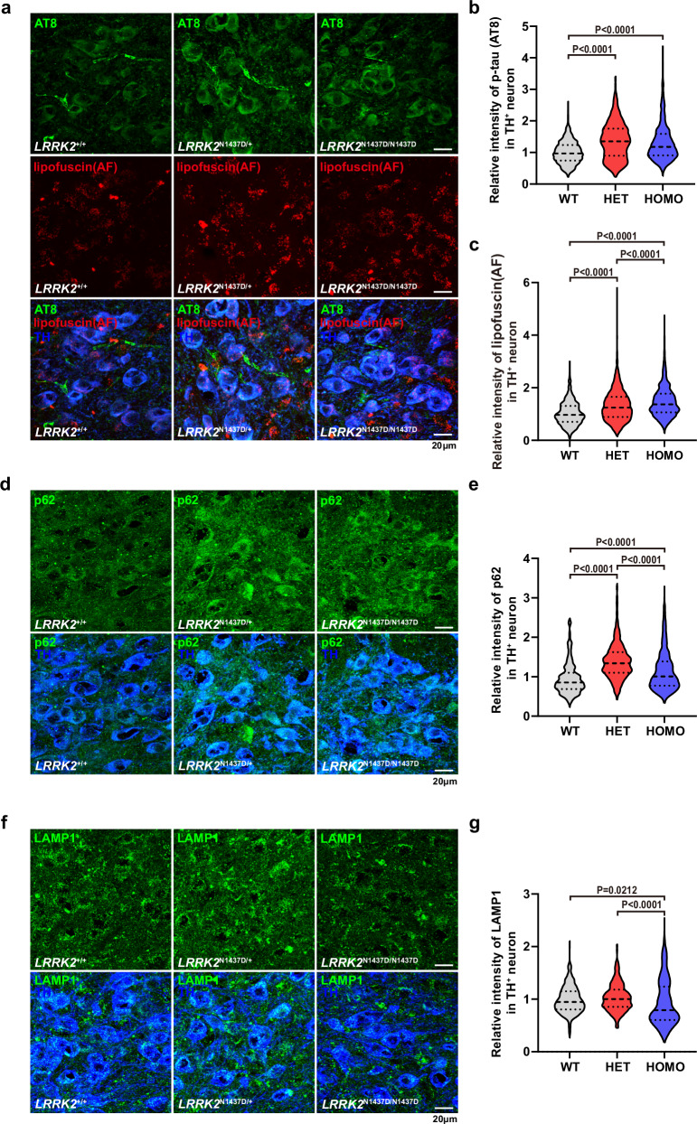
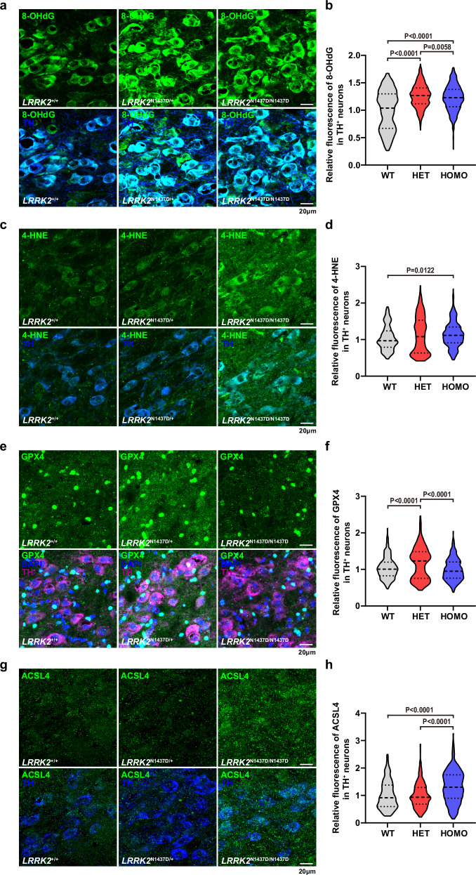
The leucine-rich repeat kinase 2 (LRRK2) gene is one of the most common genetic causes of autosomal dominant Parkinson's disease (PD) and a common genetic risk factor for sporadic PD. However, aged mice with common LRRK2 point mutations fail to exhibit age-related PD-associated behavioral and pathological impairments. We generated a novel mouse model harboring the LRRK2.p.N1437D point mutation (c.4309 A > G; NM_98578). Here, the homozygous N1437D mutation, but not the heterozygous mutation, led to an increase in the autophosphorylation, substrate phosphorylation, and GTP-binding capacity of LRRK2. Heterozygous N1437D mice also showed unaffected behavior and pathology while the homozygous mice exhibited PD-associated behavioral change at 25-26 months, dopamine system damage, lipofuscin accumulation, and lipid peroxidation in substantia nigra dopaminergic neurons at 26-27 months. The new N1437D point mutation mouse does not require LRRK2 overexpression and may better mimic the pathological characteristics of LRRK2 mutation in the ROC-COR region.


Conflict of interest statement

Competing interests: The authors declare no competing interests

Figures

Fig. 1
Fig. 1. The construction of LRRK2.p.N1437D point mutant mice.
a The illustration of the N1437D mouse construction by CRISPR/Cas9. b The breeding strategies of WT, HET, and HOMO mice used in experiments and their corresponding genotype results. The image of the mouse originates from an unlicensed cartoon depiction available on the internet, commonly utilized to illustrate mouse patterns in scientific research articles. c LRRK2 abundance from the cortex of 26–27 months in WT, HET, and HOMO mice. d N1437D mutation did not alter LRRK2 protein abundance (n = 3 for WT, n = 4 for HET, and n = 4 for HOMO). e IP enrichment test for LRRK2 from 26-27 months mouse lung. f The autophosphorylation and the GTP binding ability of N1437D mutated LRRK2 in 26-27 months mouse lung tissue. g Autophosphorylation of LRRK2 was increased in N1437D HOMO mice (Each group had three technical replicates). h The LRRK2 from 26-27 months N1437D HOMO mice lung had a stronger GTP-binding ability than the WT LRRK2 (Each group had three technical replicates). i The level of substrate phosphorylation of LRRK2 in 26–27 months lung tissue. j The N1437D mutation did not change the rab10 protein abundance (n = 4 for WT, n = 6 for HET, and n = 4 for HOMO). k The HOMO mice exhibited an increase in phosphorylation of rab10 at Thr 73 in 26-27 M lung tissue. (n = 4 for WT, n = 6 for HET, and n = 4 for HOMO). l Weight recording from 2 months to 25-26 months (2M-12M: n = 12 for WT, n = 15 for HET, and n = 14 for HOMO; 17-18 M: n = 11 for WT, n = 15 for HET, n = 13 for HOMO; 25-26 M: n = 11 for WT, n = 18 for HET, and n = 13 for HOMO). (m) Percentage of body weight loss at 17-18 months compared with 12 months (n = 11 for WT, n = 15 for HET, and n = 13 for HOMO). The uncropped blot images of this figure are listed in Fig. S8. Data are represented as mean ± SEM. * p < 0.05, **p < 0.01.
Fig. 2
Fig. 2. The N1437D mutation impaired motor performance of HOMO mice at 25-26 months of age.
a Example locomotor trajectories of 17-18 M group and 25-26 M group during the first 10 minutes of open field test. b Time spent in the central area of the open field during the first 10 min (17-18 M group: n = 12 for WT, n = 14 for HET, and n = 11 for HOMO; 25-26 M group: n = 6 for WT, n = 10 for HET, and n = 7 for HOMO). c The distance moved in 30 minutes open field test (17-18 M group: n = 12 for WT, n = 14 for HET, and n = 11 for HOMO; 25-26 M group: n = 6 for WT, n = 10 for HET, and n = 7 for HOMO). d The latency to fall in rotarod test (17-18 M group: n = 11 for WT, n = 15 for HET, and n = 13 for HOMO; 25-26 M group: n = 11 for WT, n = 17 for HET, and n = 11 for HOMO). The turn over time (e) and total time (f) spent in pole test (17-18 M group: n = 12 for WT, n = 14 for HET, and n = 11 for HOMO; 25-26 M group: n = 10 for WT, n = 17 for HET, and n = 14 for HOMO). g The grip strength of HOMO mice was significantly lower than that of WT mice at 25-26 months (n = 11 for WT, n = 18 for HET, and n = 13 for HOMO). h Example of paw print during gait analysis. i, j The print length in walking during gait analysis (17-18 M group: n = 11 for WT, n = 10 for HET, and n = 11 for HOMO; 25-26 M group: n = 6 for WT, n = 8 for HET, and n = 7 for HOMO). k, l The stride length in walking during gait analysis (17-18 M group: n = 11 for WT, n = 10 for HET, and n = 11 for HOMO; 25-26 M group: n = 6 for WT, n = 8 for HET, and n = 7 for HOMO). Right front (RF), right hind (RH), left front (LF), left hind (LH). Data are represented as mean ± SEM. * p < 0.05.
Fig. 3
Fig. 3. The N1437D mutation impaired the SN dopamine system in HOMO mice at aged 26-27 months.
Example results of 11C-CFT DAT-PET for 17-18 M mice (a) and 26-27 M mice (b), the color bar was 0-100 kBq/cc. The Relative SUVR of striatum to cerebellar for 17-18 M mice (c) and 26-27 M mice (d) in 11C-CFT DAT-PET (17-18 M group: n = 5 for WT, n = 5 for HET, and n = 6 for HOMO; 25-26 M group: n = 4 for WT, n = 5 for HET, and n = 4 for HOMO). Example of immunofluorescence staining (e) and counting (f) of TH neurons at 17-18 months in the unilateral substantia nigra (n = 5 for WT, n = 5 for HET, and n = 5 for HOMO). Example of immunofluorescence staining (g) and counting (h) of TH neurons at 26-27 months in the unilateral substantia nigra (n = 6 for WT, n = 9 for HET, and n = 8 for HOMO). Data are represented as mean ± SEM. *p< 0.05.
Fig. 4
Fig. 4. The phosphorylated tau, lipofuscin, p62, and LAMP1 levels in SN TH neurons of 26-27 months N1437D mice.
a Example of phosphorylated tau levels (AT8) and lipofuscin autofluorescence in TH neurons of substantia nigra in 26-27 months mice. b The phosphorylated tau in TH neurons of HET and HOMO mice were higher than those in WT group, but there was no significant difference between HET and HOMO mice (n = 870 neurons from 6 WT mice, n = 1259 neurons from 8 HET mice, and n = 1107 neurons from 9 HOMO mice). c The level of lipofuscin autofluorescence in TH neurons of substantia nigra was the highest in HOMO mice, followed by HET and the lowest in WT (n = 870 neurons from 6 WT mice, n = 1259 neurons from 8 HET mice, and n = 1107 neurons from 9 HOMO mice). d Example of p62 staining in TH neurons of substantia nigra in 26-27 months mice. e The p62 level in TH neurons of HET and HOMO mice were higher than those in WT mice (n = 570 neurons from 3 WT mice, n = 1064 neurons from 5 HET mice, and n = 864 neurons from 4 HOMO mice). f Example of LAMP1 staining in TH neurons of substantia nigra in 26-27 months mice. g The relative LAMP1 intensity in TH neurons of HOMO mice was significantly weaker than that of WT and HET mice (n = 460 neurons from 3 WT mice, n = 629 neurons from 6 HET mice, and n = 571 neurons from 4 HOMO mice). Data are represented as violin plot. *p < 0.05, **p < 0.01, ***p < 0.001.
Fig. 5
Fig. 5. The HOMO mice displayed oxidative damage and lipid peroxidation at 26-27 months age.
a Example of 8-OHdG staining in TH neurons of substantia nigra in 26-27 months mice. b The 8-OHdG level in TH neurons indicated HET and HOMO mice harbored higher oxidative damage compared with WT mice, (n = 478 neurons from 3 WT mice, n = 1020 neurons from 5 HET mice, and n = 625 neurons from 4 HOMO mice). c Immunofluorescence staining of 4-HNE in substantia nigra TH neurons of 26-27 months mice. d HOMO mice had higher 4-HNE level compared with WT and HET mice (n = 204 neurons from 3 WT mice, n = 294 neurons from 3 HET mice, and n = 422 neurons from 3 HOMO mice). e Example of GPX4 in TH neurons of substantia nigra in 26-27 months mice. f Among the three genotypes, HET mice had the highest GPX4 level (n = 408 neurons from 3 WT mice, n = 314 neurons from 3 HET mice, and n = 408 neurons from 3 HOMO mice). g The ACSL4 immunofluorescence in substantia nigra TH neurons of 26-27 months mice. h HOMO mice harbored higher ACSL4 level in substantia nigra TH neurons (n = 191 neurons from 3 WT mice, n = 451 neurons from 5 HET mice, and n = 378 neurons from 4 HOMO mice). Data are represented as violin plot. *p < 0.05, **p < 0.01, ***p < 0.001.
Fig. 6
Fig. 6. The potential pathogenic mechanism of N1437D mutation.
The results of immunofluorescence showed that N1437D mutation induced increased oxidative damage in TH neurons of HET and HOMO elder mice. However, for HET mice, the presence of anti-lipid peroxidation GPX4 conferred resistance against the enhancive oxidative damage caused by the N1437D mutation, resulting in no significant differences of behavior and pathology between HET and WT mice. Conversely, in HOMO mice, there was a hindered increase in lipid-protecting GPX4 levels while lipid peroxidation ACSL4 levels rose. These findings suggest that lipid peroxidation may be implicated as a causative factor for SN dopamine system damage and motor function degeneration observed in N1437D HOMO mice. We appreciate Mr. Kai Qian for drawing the pictures of aged mice and aged tremor mice.

References

    1. Aasly, J. O. et al. Novel pathogenic LRRK2 p.Asn1437His substitution in familial Parkinson’s disease. Mov. Disord.25, 2156–2163 (2010). - PMC - PubMed
    1. Alcalay, R. N. et al. Cognitive and motor function in long-duration PARKIN-associated Parkinson disease. JAMA Neurol.71, 62–67 (2014). - PMC - PubMed
    1. Alessi, D. R. & Sammler, E. LRRK2 kinase in Parkinson’s disease. Science360, 36–37 (2018). - PubMed
    1. Yue, M. et al. Progressive dopaminergic alterations and mitochondrial abnormalities in LRRK2 G2019S knock-in mice. Neurobiol. Dis.78, 172–195 (2015). - PMC - PubMed
    1. Tong, Y. et al. R1441C mutation in LRRK2 impairs dopaminergic neurotransmission in mice. Proc. Natl. Acad. Sci. USA106, 14622–14627 (2009). - PMC - PubMed

LinkOut - more resources