

Biomechanical effects of fascial hydrorelease: a cadaveric study
- PMID: 40155875
- PMCID: PMC11951565
- DOI: 10.1186/s12891-025-08533-y
Biomechanical effects of fascial hydrorelease: a cadaveric study
Erratum in
-
Correction: Biomechanical effects of fascial hydrorelease: a cadaveric study.BMC Musculoskelet Disord. 2025 Jun 30;26(1):579. doi: 10.1186/s12891-025-08885-5. BMC Musculoskelet Disord. 2025. PMID: 40588752 Free PMC article. No abstract available.
Abstract
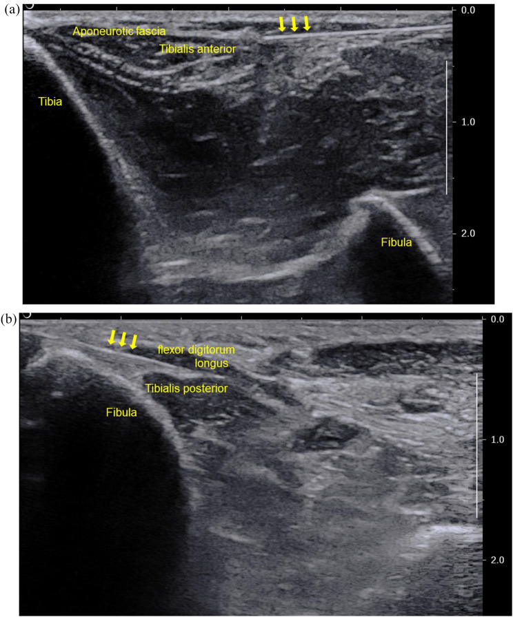
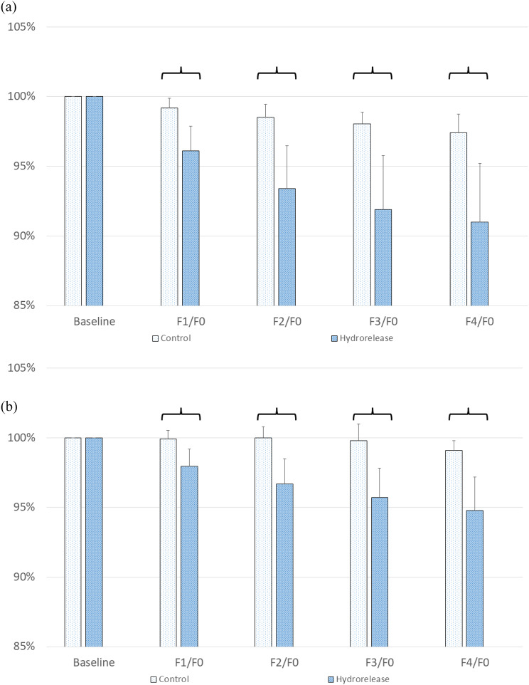
Background: We aimed to investigate the influence of hydrorelease (HR) on the gliding resistance force between the aponeurotic fascia and epimysial fascia of tibialis anterior and between two epimysial fasciae of tibialis posterior and flexor digitorum longus using a biomechanical testing system.
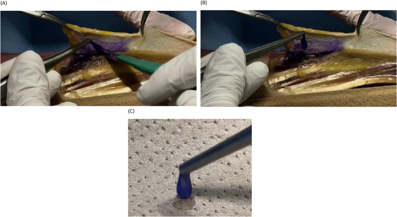
Methods: In this cadaveric comparative study, 12 paired legs amputated above the knee joint from six fresh-frozen specimens were divided into two groups. The distal insertions of the target tendons of the tibialis anterior and posterior were detached and sutured to a force gauge for tension measurement during tendon pull. These tendons were representatives of the layer between the aponeurotic and epimysial fasciae of the tibialis anterior and between the epimysial fasciae of the tibialis posterior and flexor digitorum longus. For the baseline, the position where the tension of the target tendon was approximately 15 N was determined to eliminate creep. In the HR group, the baseline test position was replicated, and force was measured. The intervention was an HR injection between the specified fascial layers. The main outcome was the gliding resistance force between the aponeurotic and epimysial fasciae and between two epimysial fasciae.
Results: The resistance force between the aponeurotic and epimysial fasciae in the HR group was 6.4% lower than that in the control group (P = 0.02). The resistance force between two epimysial fasciae in the HR group was 4.3% lower than that in the control group (P < 0.01).
Conclusions: The gliding resistance force significantly decreased after HR in the layer between the aponeurotic and epimysial fasciae and between two epimysial fasciae in this cadaveric study.
Keywords: Biomechanics; Cadaver; Fascia; Myofascial pain syndrome; Tendon.
© 2025. The Author(s).
Conflict of interest statement
Declarations. Ethics approval and consent to participate: The study was approved by the Sapporo Medical University Ethics Board (number 1-2-68). All participants provided written informed consent. Consent for publication: Not applicable. Competing interests: The authors declare no competing interests.
Figures

References
-
- Fukui S, Rokutanda R, Kawaai S, Suda M, Iwata F, Okada M, et al. Current evidence and practical knowledge for ultrasound-guided procedures in rheumatology: joint aspiration, injection, and other applications. Best Pract Res Clin Rheumatol. 2023;37:101832. - PubMed
-
- Neo EJR, Shan NT, Tay SS. Hydrodissection for carpal tunnel syndrome: a systematic review. Am J Phys Med Rehabil. 2022;101:530–9. - PubMed
-
- Courseault J, Kessler E, Moran A, Labbe A. Fascial hydrodissection for chronic hamstring injury. Curr Sports Med Rep. 2019;18:416–20. - PubMed
-
- Simons DG. New aspects of myofascial trigger points: etiological and clinical. J Musculoskelet Pain. 2004;12:15–21.
-
- Giamberardino MA, Tafuri E, Savini A, Fabrizio A, Affaitati G, Lerza R, et al. Contribution of myofascial trigger points to migraine symptoms. J Pain. 2007;8:869–78. - PubMed
Publication types
MeSH terms
Grants and funding
LinkOut - more resources
Full Text Sources
