. 2025 Mar 28;26(1):75.
doi: 10.1186/s13059-025-03521-w.

Pamoic acid and carbenoxolone specifically inhibit CRISPR/Cas9 in bacteria, mammalian cells, and mice in a DNA topology-specific manner

Affiliations

Pamoic acid and carbenoxolone specifically inhibit CRISPR/Cas9 in bacteria, mammalian cells, and mice in a DNA topology-specific manner

Yuxuan Zhang et al. Genome Biol. .

Abstract

Background: Regulation of the target DNA cleavage activity of CRISPR/Cas has naturally evolved in a few bacteria or bacteriophages but is lacking in higher species. Thus, identification of bioactive agents and mechanisms that can suppress the activity of Cas9 is urgently needed to rebalance this new genetic pressure.

Results: Here, we identify four specific inhibitors of Cas9 by screening 4607 compounds that could inhibit the endonuclease activity of Cas9 via three distinct mechanisms: substrate-competitive and protospacer adjacent motif (PAM)-binding site-occupation; substrate-targeting; and sgRNA-targeting mechanisms. These inhibitors inhibit, in a dose-dependent manner, the activity of Streptococcus pyogenes Cas9 (SpyCas9), Staphylococcus aureus Cas9 (SauCas9), and SpyCas9 nickase-based BE4 base editors in in vitro purified enzyme assays, bacteria, mammalian cells, and mice. Importantly, pamoic acid and carbenoxolone show DNA-topology selectivity and preferentially inhibit the cleavage of linear DNA compared with a supercoiled plasmid.

Conclusions: These pharmacologically selective inhibitors and new mechanisms offer new tools for controlling the DNA-topology selective activity of Cas9.

Keywords: Anti-CRISPR; DNA topology; Mice model of hydrodynamic injection; Mode of action; Selective small-molecule inhibitors.


Conflict of interest statement

Declarations. Ethics approval and consent to participate: The animal study was approved by the Institutional Animal Care and Use Committee (IACUC) of Shanghai Jiao Tong University, China. Consent for publication: Not applicable. Competing interests: The authors declare no competing interests.

Figures

Fig. 1
Fig. 1
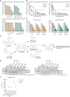
The primary inhibitor screening and identification of FDA-approved drugs to inhibit the endonuclease activity of Cas9 with a purified enzyme-based activity assay. A The primary inhibitor screening for suppressing the endonuclease activity of SpyCas9 and the schematic diagram for the in vitro enzyme assay, bacteria, mammalian cells, or animal -based studies of Cas9 inhibitors. After a drug screening of 4607 compounds at 200 μM in the activity assay of Cas9, 31 compounds were found to dose-dependently inhibit the activity of Cas9 with IC50 values less than 200 μM. Five reprensentative leads were selected for the efficacy and selectivity studies on purified enzyme, bacteria, mammalian cell or animal levels. B The effect of five leads as well as AcrIIA4 on the cleavage activity of SpyCas9 to the circular plasmid. Pamoic acid, epirubicin, dalbavancin, carbenoxolone or docusate at the indicated concentrations was incubated with a complex of SpyCas9 (375 nM) and sgRNA (400 nM) and tested under the standard assay conditions (Additional file 1:Fig.S1B-C;see the “Methods” section). The pcDNA3.1/CT-GFP plasmid (300 ng) containing a protospacer sequence (N20: 5’-CCAATTCTTGTTGAATTAGA-3’) and a 5’-TGG PAM (Additional file 1: Table S4) was used in the assay. AcrIIA4, a known anti-CRISPR protein inhibitor of Cas9 [22], was also tested under the standard assay conditions with an exception of using 22 °C for the assay instead of 37 °C, which was reported for AcrIIA4 to show a better inhibition to SpyCas9 [23]. In TBE buffer, the linear form of pcDNA3.1/CT-GFP plasmid (~ 7200 bp) migrates faster than the covalently closed circular DNA (cccDNA) form of the plasmid, which was reported previously [21] and also validated by us (Additional file 1:Fig.S1B).Means ± SDs (n = 3, biological replicates). C The effect of five leads as well as AcrIIA4 on the cleavage activity of SpyCas9 to the linear oligonucleotide substrate. Pamoic acid, epirubicin, dalbavancin, carbenoxolone, or docusate as well as AcrIIA4 was tested under the assay conditions described above (B) using a FAM-labeled 87 bp oligonucleotide as the substrate (10 nM; linear substrate, thereafter), which bears the same sequence of protospacer and PAM to the pcDNA3.1/CT-GFP plasmid (B). Eighty seven base pairs, the 5′-FAM-labeled substrate. Thirty base pairs, the FAM-labeled cleaved 5′-terminal fragment. Means ± SDs (n = 3, biological replicates). D,E The effect of five leads on the enzymatic activity of SauCas9. Pamoic, epirubicin, dalbavancin, carbenoxolone, or docusate at the indicated concentrations were incubated with the complex of SauCas9 (100 nM) and sgRNA (100 nM) before the addition of the pcDNA3.1/CT-GFP plasmid (300 ng, D) or a FAM-labeled 87 bp oligonucleotide (10 nM, E). The two forms of DNA substrate of this assay share the same sequence of protospacer (N21, 5′-CTGGAGTTGTCCCAATTCTTG-3′) and 5′-TTGAAT PAM (Additional file 1: Table S4). The original images for circular plasmid and linear oligonucleotide are shown in the Additional file 1: Fig. S3F and S3G, respectively. Means ± SDs (n = 3, biological replicates). DNA only, pcDNA3.1/CT-GFP plasmid (B), or 87 bp linear DNA (C) in the presence of assay buffer (see the “Methods” section). The optical density of the DNA band for the linear form (agarose gels, B) or for the 30 bp product (polyacrylamide gels, C), as well as the area containing the linear, open circular, and supercoil bands of the plasmid (B) or the area containing the 87 bp and 30 bp products (C) was quantified using ImageJ software (NIH, Bethesda, MD). The cleavage activity of Cas9 was calculated by dividing the band density of the linear form (B) or 30 bp product (C) by the density of total area in the same lane (100%) and is shown as a percentage below the gel. The cleavage activity was normalized to that of the DMSO group (100%), as shown in the right panel. Statistical analysis was performed using one-way ANOVA with Tukey’s multiple comparisons test; *p < 0.05; **p < 0.01; ***p < 0.001. The experiments were independently repeated three times, and representative results are presented
Fig. 2
Fig. 2
The biochemical mechanisms of five representative leads on inhibiting SpyCas9. A Surface plasmon resonance assay analysis of the binding of five representative leads to SpyCas9. SpyCas9 at a concentration of 100 μg/mL was immobilized onto the surface of the CM5 sensor chip and then employed to analyse the binding of pamoic acid, dalbavancin or carbenoxolone as well as epirubicin and docusate (Additional file 1:Fig. S4D). The equilibrium affinity constant (KD) values were calculated with BIAcore evaluation software (version 3.1). B The pre-incubation of pamoic acid or carbenoxolone, but not dalbavancin, epirubicin or docusate, with SpyCas9 ribonucleoprotein enhances their inhibitory effects on the activity of Cas9. Pamoic acid or carbenoxolone at the indicated concentrations were pre-incubated with Cas9: sgRNA (375 nM: 400 nM) for 15 min before adding the GFP plasmid (condition a of left panel), or exposed to the enzyme together with the plasmid without a pre-incubation (condition b). Then, the samples were tested under the standard assay conditions. Means ± SDs (n = 3, technical replicates). The results for other inhibitors were slowed in the Additional file 1: Fig. S4E. C-D Pamoic acid or carbenoxolone, but not dalbavancin, docusate or epirubicin, specifically prevents the binding of DNA substrate to SpyCas9 in DNA EMSA assays. dCas9 in the presence of sgRNA (C or right panelof D) or in the absence (left panelof D) was incubated with the 5’-FAM labeled linear DNA substrate or plasmid and the inhibitor under the indicated sampling order, before a separation on a 6% native polyacrylamide gel (upper panel of C for the 87 bp oligonucleotide; see the “Methods” section) or a 1% native agarose gel (lower panel of C for the plasmid). The polyacrylamide gels were stained with 1 × SYBR™ Gold (the right panel of C and D) or were imaged for fluroresence of FAM under an excitation of 488 nm on a ChemiDoc scanner (left and upper panels of C; left panel of D), while the agarose gel was dying with EB and illuminated under UV (lower panels of C). The lower and right panel of C, the plasmid DNA (the lower and left panel) treated with the indicated compounds in the absence of dCas9 and sgRNA. E Dalbavancin binds to free sgRNA or dCas9: sgRNA complex in a sgRNA EMSA assay. The free sgRNA (left panel) or sgRNA captured with dCas9 (right panel) was incubated with the inhibitor before a staining with 1 × SYBR™ Gold. F Epirubicin as well as doxorubicin or cisplatin directly binds to free DNA substrates. The plasmid (300 ng, equal to 5 nM) or oligonucleotide (10 nM) was pre-incubated with the compounds before adding of 5 × loading buffer containing 1.2% SDS. The samples were heated for 5 min at 95 °C and separated on respective gels (see above). G Oligonucleotide containing 8 × PAM rescues the destabilization of pamoic acid or carbenoxolone on Cas9 ribonucleoprotein in a thermal shift assay. The complex of Cas9 and sgRNA at 1 μM was incubated with 200 μM pamoic acid, 100 μM carbenoxolone or 50 μM dalbavancin for 15 min in the presence of the oligonucleotide containing of 8 × PAM at a molar ratio of 0.5:1, 1:1, 2:1 or 5:1 to Cas9 (see the “Methods” section). The melting curves of Cas9 were showed in the Additional file 1:Fig.S5D and the unfolding transition midpoint temperature (Tm) of Cas9 was calculated by the GraphPad software (version 8.4, San Diego, CA). Means ± SDs (n = 3, biological replicates). Statistical analysis was performed using one-way ANOVA with Tukey’s multiple comparisons test; ns, no significance; **p < 0.01; ***p < 0.001. H The dose-dependent inhibitory effect of compounds on the cleavage of target strand ssDNA by Cas9. The activity assay was performed using 5’-FAM labeled 87 bp single stand oligonucleotide that is reverse and complement to the anti-GFP sgRNA and without the sequence of PAM (Additional file 1: Table S3-4), instead of the annealed double strand oligonucleotide. The original image was shown in the Additional file 1:Fig.S5F and the normalized data are shown here. Means ± SDs (n = 3, technical replicates). DNA only, pcDNA3.1/CT-GFP plasmid (B, lower panel of C and upper panel of F), or 87 bp linear DNA (upper panel of C, D and lower panel of F) in the presence of assay buffer. The cleavage activity of Cas9 was determined and is shown as a percentage of the DMSO group (100%; right panel of B or H). All experiments were independently repeated at least twice, and representative results are presented
Fig. 3
Fig. 3
The selectivity and structure–activity relationship of Cas9 inhibitor. A The effect of pamoic acid or carbenoxolone on inhibiting the activity of Cas9-VQR (upper) or SpRY-Cas9 (lower) mutant. Compounds were tested under the standard conditions with using the Cas9-VQR (D1135V/R1335Q/T1337R) or SpRY-Cas9 (A61R/L1111R/D1135L/S1136W/G1218K/E1219Q/N1317R/A1322R/R1333P/R1335Q/T1337R; NEB, M0669T) and the FAM-labeled 87 bp GFP oligonucleotide with a 5’-NGA or 5’-NAG PAM (Additional file 1: Table S2 and S4) [61], the latter of which is the preferred 5’-NHN PAM (H = A, C, or T) for SpRY-Cas9. The original images are shown in the Additional file 1: Fig. S6B. Means ± SDs (n = 3, biological replicates). B The effect of pamoic acid or carbenoxolone on inhibiting the cleavage of GFP plasmids containing a 5’-NAG or 5’-NGA PAM by SpyCas9. The original images are shown in the Additional file 1: Fig. S6D. Means ± SDs (n = 3, technical replicates). C The effect of pamoic acid or carbenoxolone on the activity of SpyCas9 nickase. Compounds were tested under the standard conditions using the wt SpyCas9 or nickase, i.e. Cas9 D10A or Cas9 H840A mutant, and the linear GFP substrate. The original images were shown in the Additional file 1: Fig. S6E. Means ± SDs (n = 3, biological replicates). D The structure-activity relationship of pamoic acid on inhibiting the activity of SpyCas9 (see Additional file 1:Fig. S6F for the original images). The common structural moiety between the Asp60-Glu70 dipeptide (right panel) and pamoic acid is shown in orange. E The effect of compounds on the activity of FnoCas12a or LbaCas12a, Type V-A CRISPR/Cas enzyme with a respective 5’-TTN PAM and a 5'-TTTV PAM. The commercially available FnoCas12a (left panel) or LbaCas12a (right panel) CRISPR/Cas enzymes (#32106-03 or #32108-03 of ToloBio, Shanghai, China) were incubated with indicated compounds for 15 min before loading the pre-mixture of DNA substrate (DNA methyltransferase 1 fragment) and crRNA (see the “Methods” section). The 825 bp substrate or 525 bp and 300 bp products of DNA methyltransferase 1 was separated on 1% agarose gel and stained with EB. The optical density of the DNA bands was determined with ImageJ and the cleavage activity was showed as percentages (n = 2, biological replicates; see the “Methods” section). F The effect of Cas9 inhibitors on the activity of EcoRI or KpnI endonuclease. Compounds were tested in assays containing 375 nM Cas9 or 0.5 μl EcoRI or KpnI (7.5 U for EcoRI and 10 U for KpnI per assay) and 300 ng ~2.5 kb linear DNA (Additional file 1: Fig. S3C). The original images were shown in the Additional file 1: Fig. S7A. ATA, aurintricarboxylic acid [30] (Additional file 1: Table S1 and Additional file 1: Fig. S2). Statistical analysis was performed using one-way ANOVA with Tukey’s multiple comparisons test; *p < 0.05; **p < 0.01; ***p < 0.001. All experiments were independently repeated at least twice, and representative results are presented
Fig. 4
Fig. 4
The effect of inhibitors on suppressing the genome editing on SSEA locus by SpyCas9 or SauCas9 in bacteria. A The schematic diagram of two-plasmid-based bacterial survival assay for detecting the activity of SpyCas9 in bacteria. pCas plasmid (Addgene #62225,Additional file 1: Table S2) was transformed into the E. coli MG1655 before an electroporation with pTarget plasmids coding for sgRNA targeting SSEA (pT-sgSSEA, Additional file 1: Table S4) or empty vector (pT-sgControl; pTargetF, Addgene #62226). Then, the effect of compounds was evaluated by counting the number of survival colonies (see the “Methods” section). B The effect of Cas9 inhibitors on the activity of SpyCas9 in the two-plasmid-based bacterial survival assay. Compounds were incubated with the electroporated bacteria (A) for 1.5 h at 32 °C before spreading on LB plates with kanamycin and spectinomycin antibiotics. After an overnight incubation, the image of cultured plate was taken (Additional file 1:Fig.S7D), and the number of colonies (indicated below the plate image) was quantified with a Colony Counter software (Tanon, China), and expressed as the percentages of their respective control at the same concentration (the pT-sgControl electroporated strain treated with the compound, 100%; Additional file 1:Fig.S7D). Means ± SDs (n = 3, biological replicates). Statistical analyses were performed using two-way ANOVA with Bonferroni posttests; *p < 0.05; **p < 0.01; ***p < 0.001. C The schematic diagram of the genome editing assay for monitoring the activity of SpyCas9 or SauCas9 in the presence of homologous repair template DNA and in bacteria. pCas plasmid was first transformed into MG1655 strain before an electroporation to pTarget plasmids carrying a pair of homologous repair template DNA that is missing the 1-243 bp of SSEA gene (see the “Methods” section) and a coding sequence for SpyCas9 or SauCas9 sgRNA (pT-sgSSEA(867 bp)). Then, the E. coli cells were incubated with the compounds before genotyping with PCR for analyzing the efficiency of genome editing at the SSEA. 909 bp, the size of PCR product from a strain, in which 1-243 bp of SSEA is missing; 1152 bp, the size of PCR product from a strain with wt SSEA gene. D The effect of Cas9 inhibitors on the genome editing activity of SpyCas9 (left panel) or SauCas9 (right panel) in the presence of a homologous recombination repair template. Compounds were incubated with the electroporated bacteria (C) for 20 h at 32 °C, and the SSEA in the treated bacteria was amplified with PCR and analyzed on a 1% agarose. The original images for PCR-based genome typing were shown in the Additional file 1:Fig.S8B. Means ± SDs (n = 3, biological replicates). The ddH2O (for dalbavancin or carbenoxolone) or DMSO (pamoic acid or epirubicin) treated groups, 100%. Statistical analysis was performed using one-way ANOVA with Tukey’s multiple comparisons test; ***p < 0.001. All experiments were independently repeated at least twice, and representative results are presented
Fig. 5
Fig. 5
The effect of inhibitors on suppressing the editing of exogenous GFP gene, endogenous COSMC or EMX1 loci by SpyCas9, SauCas9 or BE4 in HEK293FT cells. A The schematic diagram of cellular assays for analyzing the effects of inhibitors on the activity of SpyCas9, SauCas9 or BE4 for the cleavage of GFP, COSMC or EMX1 gene in HEK293FT cells. The cells were transfected with Lipofectamine™ 3000 Transfection Reagent (Thermal Fisher, L3000008) in the presence of plasmids containing SpyCas9, SauCas9 or their sgRNAs before an incubation with the compound for 24 h. After the colony selection with puromycin, genomic DNA of the cells was then extracted, and the target gene was amplified with PCR (see the “Methods” section). The editing efficiency of Cas9 was analyzed with flow cytometry, TIDE, EditR or T7EI method [–68]. B Flow cytometry analysis of the effect of compounds on the disruption of GFP by SpyCas9 in HEK293FT cells. The cells stably expressing of GFP were transiently transfected with the PX459-SpyCas9 (Addgene #62988) and pGL3 plasmids coding anti-GFP sgRNA (the blue line) or control sgRNA (the green line) before an incubation with the compound for 24 h (A). The GFP fluorescent from the treated cells was analyzed with LSR Fortessa flow cytometer (BD Biosciences, Franklin Lakes, NJ), and the gating strategy and corresponding histograms are shown in the Additional file 1:Fig.S9A-B. The quantitative data was shown as the percentage of the group treated with control sgRNA (100%). Means ± SDs (n = 3, biological replicates). C The effect of compounds on the editing of endogenous COSMC or EMX1 by SpyCas9 in HEK293FT wt cells. The cells were transfected with PX459-SpyCas9 plasmid coding anti-COSMC, anti-EMX1 sgRNA (for the spacer sequences, see Additional file 1: Table S4) or control sgRNA (the SpyCas9 only group, Additional file 1:Fig.S9D),before the treatment with the indicated compounds for 24 h (A). The target DNA was accordingly amplified with PCR before subjected to Sanger sequencing (Additional file 1:Fig.S9E-Gand Additional file 1:Fig.S10A-C). The editing efficiency of Cas9 was analyzed with the TIDE method (https://tide.nki.nl/), normalized with the DMSO (for pamoic acid; final concentrations, 0.4%) or ddH2O (for dalbavancin or carbenoxolone) -treated groups (100%) and shown. AcrIIA4 was used as a positive control (Additional file 1:Fig.S10D). Means ± SDs (n = 3, biological replicates). D T7EI mismatch detection assay analysis of the inhibitory effect of compounds on editing of COSMC by SpyCas9 in HEK293FT cells. The amplified PCR products (541 bp; input) from the extracted genomic DNA of the control (the left four lanes of each gel) or edited HEK293FT cells (C), which have been treated with DMSO or ddH2O (the middle four lanes), 100 μM pamoic acid, 50 μM dalbavancin or 50 μM carbenoxolone (the right four lanes), were incubated with T7EI or without the treatment at 37 °C for 15 min before an analysis the COSMC site with 2% agarose gels (see the “Methods” section). The percentage of Indel was calculated by the formula dividing the band density of fragments to that of total input and normalized with the background value [69]. E The effect of compounds on the base editing of EMX1 by BE4 base editor in HEK293FT cells. The cells were co-transfected with the BE4 and pGL3 plasmids, the latter coding an anti-EMX1 sgRNA that is the same as the one used for SpyCas9 (Additional file 1: Table S4), and treated with the compounds for 24 h (A). The amplified PCR products from the EMX1 site were accordingly sequenced (Additional file 1:Fig.S11E-F), and the converting rates of C5 or C6 base in the N20 sequence were analyzed with EditR (https://moriaritylab.shinyapps.io/editr_v10/;upperpanels) [66, 67]. The converting rate at the C5 and C6 position were normalized with the respective control (100%) and the base editing efficiency was shown as percentages for both the C5 and C6 sites (lower panels). Means ± SDs (n = 3, biological replicates). F The effect of compounds on the editing of COSMC by SauCas9 in HEK293FT cells. The cells were co-transfected with pX601-SauCas9 (Addgene #107055) and pGL3 plasmids containing an anti-COSMC sgRNA (Additional file 1: Table S2 and S4), before the treatment with the compounds for 24 h (A). The editing efficiency of Cas9 was analyzed with the TIDE method (https://tide.nki.nl/) and presented as percentages of control (100%). The raw sequencing results were shown in the Additional file 1:Fig.S12A-C. Means ± SDs (n = 3, biological replicates). G The comparison for the effects of pamoic acid or carbenoxolone on the SpyCas9-mediated disruption of GFP plasmids with a 5’-NGG or 5’-NAG PAM. The HEK293FT wt cells were transiently co-transfected with the PX459-SpyCas9 and pCDH-GFP plasmids, which contains a 5’-NGG PAM or an in situ mutated 5’-NAG PAM (see the “Methods” section) and were treated with the compounds 6 h post the transfection and for 24 h. The GFP fluorescent from the cells was accordingly quantified with flow cytometer and the data was shown as presentages of the control (100%; for the raw histograms, see Additional file 1:Fig.S12E; see the “Methods” section). Means ± SDs (n = 6, biological replicates). Statistical analysis was performed using one-way ANOVA with Tukey’s multiple comparisons test (B, C, E or F) or two-way ANOVA with Bonferroni posttests (G). *p < 0.05, **p < 0.01, ***p < 0.001. All experiments were independently repeated at least twice, and representative results are presented
Fig. 6
Fig. 6
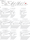
The effects of inhibitors on antagonizing the Cas9-mediated genome editing in a hydrodynamic-injection-based anti-PCSK9 mice model. A Scheme of experiments for the administration of compounds and construction of a SpyCas9-mediated anti-PCSK9 mouse model by hydrodynamic injection. BALB/c mice were intraperitoneally injected with the compounds at doses of 10, 20, 40, 80 or 160 mg/kg on day 0, followed by a hydrodynamic injection of 90 mμg of PX459-SpyCas9 plasmids encoding anti-PCSK9 sgRNA (Additional file 1: Table S4) [76] or control (vehicle group), and 5-8 s into the tail vein. Blood was collected via orbital bleeding on day 2. Two additional cycles of intraperitoneal injection of compounds (at the same dose as the 1st treatment), hydrodynamic injection of PX459-SpyCas9 plasmids, or orbital bleeding to mice were performed on days 3-8 before the mice were sacrificed on day 8. B Pamoic acid, dalbavancin or carbenoxolone restored the circulatory amount of PCSK9 in a SpyCas9-mediated anti-PCSK9 mouse model. After collecting the blood sample from the orbital sinus on Day 2 (1st orbital blood collection), Day 5 (2nd orbital blood collection) or Day 8 (3rd orbital blood collection), the amount of plasma PCSK9 was determined by Mouse Proprotein Convertase 9/PCSK9 Quantikine ELISA Kit (R&D, MPC900, Minneapolis, MN). Pamoic acid and dalbavancin were studied together in experiment 1, whereas carbenoxolone was studied in experiment 2. Means ± SDs (the groups of Cas9 + sgPCSK9 + saline and + 80 mg/kg pamoic acid in the 1st and 2nd treatment cycles of experiment 1, n = 12; the other groups, n = 6). Statistical analysis was performed using one-way ANOVA with Dunnett’s multiple comparisons test (for the samples from 1st and 2nd treatment cycles of experiment 1) or with Tukey’s multiple comparisons test (for the samples from the 3rd treatment cycles of experiment 1 and the samples from experiment 2). Ns, no significance; *p < 0.05; **p < 0.01; ***p < 0.001
Fig. 7
Fig. 7
Three proposed modes of action of four newly identified leads in inhibiting the activity of Cas9. Mode of action: pamoic acid or carbenoxolone binds competitively with DNA substrates to the PAM binding site of Cas9 positioned by Arg1333 and Arg1335, and shows a various inhibitory potency to the cleavage of the supercoil plasmid or linear DNA substrate by Cas9, and thus leading a selectivity on the cleavage of different topology of DNA. Dalbavancin directly binds to sgRNA and forms the quaternary complexes of Cas9 and sgRNA. Epirubicin directly binds to the DNA substrate and forms the quaternary complexes of Cas9 and DNA

References

    1. Jinek M, Chylinski K, Fonfara I, Hauer M, Doudna JA, Charpentier E. A programmable dual-RNA-guided DNA endonuclease in adaptive bacterial immunity. Science. 2012;337:816–21. - PMC - PubMed
    1. Cong L, Ran FA, Cox D, Lin S, Barretto R, Habib N, Hsu PD, Wu X, Jiang W, Marraffini LA, et al. Multiplex genome engineering using CRISPR/Cas systems. Science. 2013;339:819–23. - PMC - PubMed
    1. Nishimasu H, Ran FA, Hsu PD, Konermann S, Shehata SI, Dohmae N, Ishitani R, Zhang F, Nureki O. Crystal structure of Cas9 in complex with guide RNA and target DNA. Cell. 2014;156:935–49. - PMC - PubMed
    1. Anders C, Niewoehner O, Duerst A, Jinek M. Structural basis of PAM-dependent target DNA recognition by the Cas9 endonuclease. Nature. 2014;513:569–73. - PMC - PubMed
    1. Jinek M, Jiang F, Taylor DW, Sternberg SH, Kaya E, Ma E, Anders C, Hauer M, Zhou K, Lin S, et al. Structures of Cas9 endonucleases reveal RNA-mediated conformational activation. Science. 2014;343:1247997. - PMC - PubMed

LinkOut - more resources