. 2025 Mar 29;145(1):217.
doi: 10.1007/s00402-025-05759-2.

Bone defects in knee revision arthroplasty-a therapy-oriented classification

Affiliations

Bone defects in knee revision arthroplasty-a therapy-oriented classification

Max Jaenisch et al. Arch Orthop Trauma Surg. .

Abstract

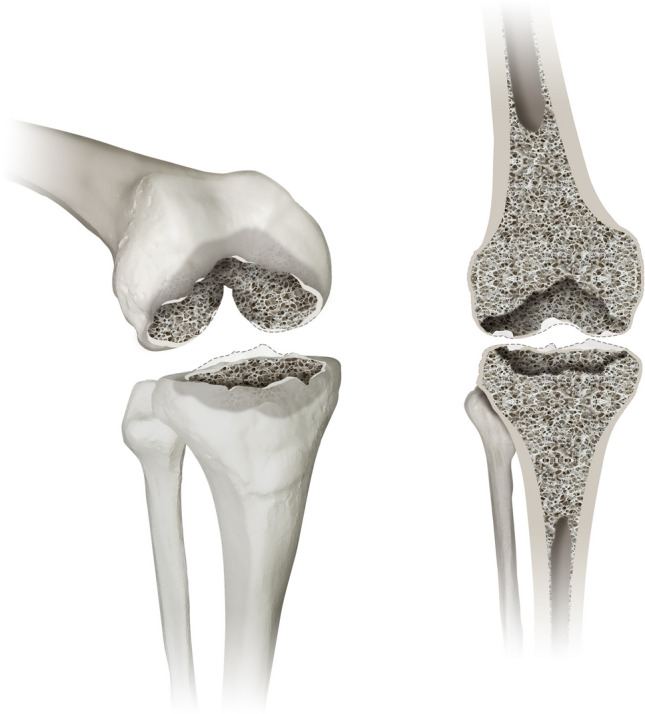
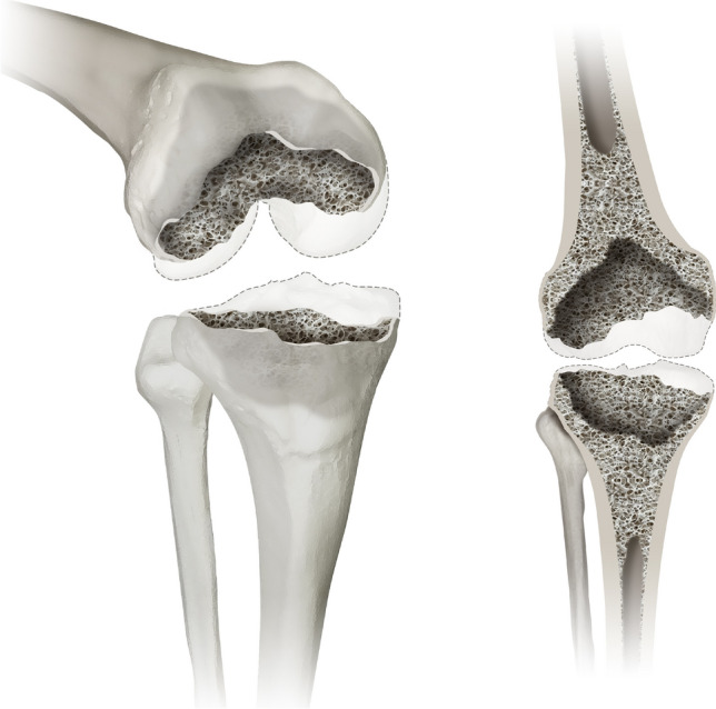
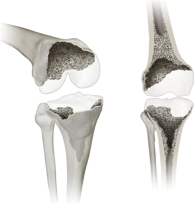
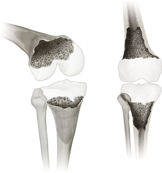
Introduction: The complex field of revision knee arthroplasty displays a lack of standardized, intuitive pre- and intraoperative assessment of bone defects. In clinical practice revision surgeries are a common sight presenting with increasingly complex cases of bone loss and ligamentary dysfunction. To address these issues the Knee Defect Classification (KDC) is introduced to offer a reliable, reproducible and an intuitive classification system with a clear therapeutic guideline.

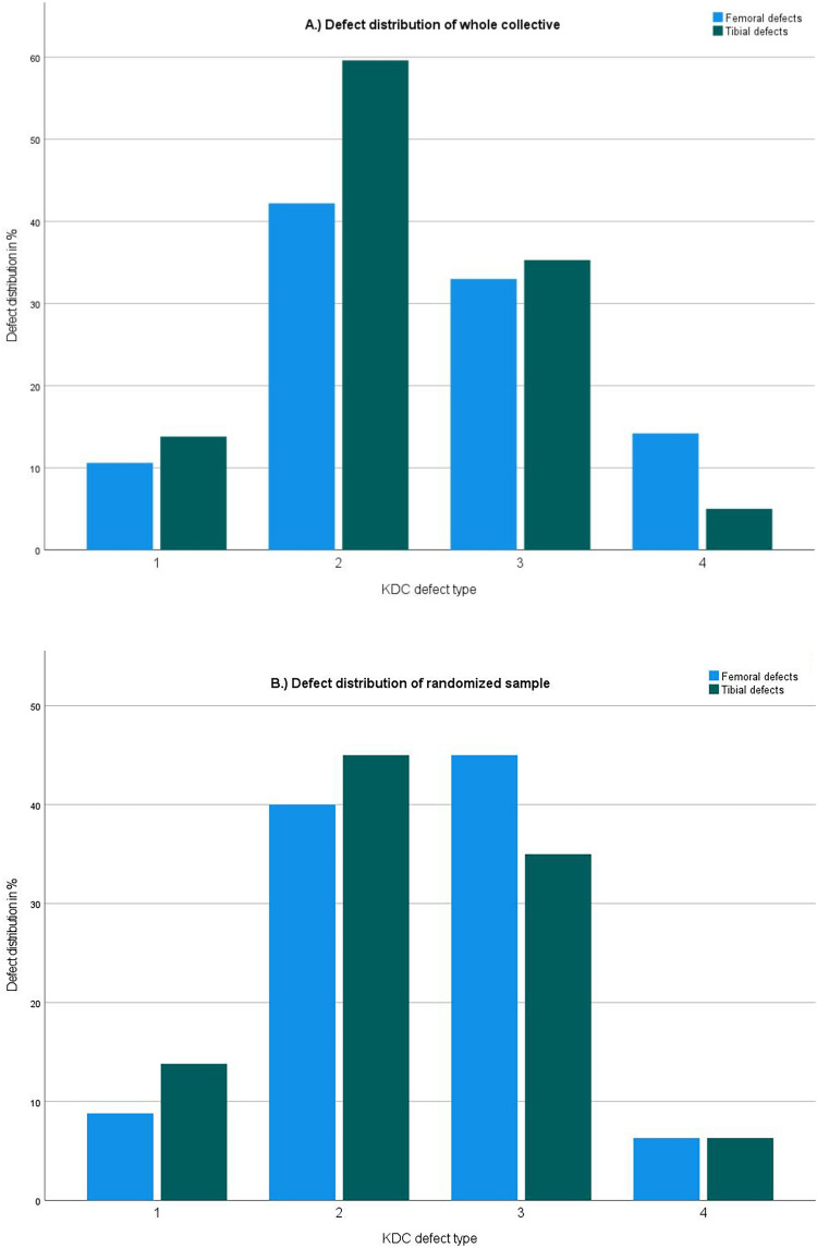
Materials and methods: Reliability was evaluated through comparison between intraoperative findings and preoperative gradings of 218 patients. To account for reproducibility inter- and intra-rater agreement was assessed.

Results: The mean agreement between intraoperative and radiographic grading of femoral and tibial defects was evaluated with a Cohens kappa of 0.79, representing a good agreement. For interrater reliability a mean Fleiss kappa of 0.71 could be evaluated for femoral and tibial defects falling into the good agreement range. In the subgroup analysis femoral defects presented with a Fleiss kappa of 0.652 ± 0.026 (good agreement), while tibial defects presented with a Fleiss kappa of 0.768 ± 0.024 (good agreement). For intrarater reliability a mean Cohens kappa was evaluated at 0.78 indicating good agreement.

Conclusion: The KDC is a reliable and reproducible classification system. Through its structured design it facilitates intuitive use and allows for consistent preoperative planning and intraoperative guidance. A therapeutic algorithm is provided based on a review of the literature in combination with expert opinion.

Keywords: AORI; Constrainment; Defect classification; Knee revision arthroplasty; Ligamentary instability; Reliability; Reproducability.


Conflict of interest statement

Declarations. Conflict of interest: All authors declare that they have no conflict of interest. The authors did not receive support from any organization for the submitted work. Ethical approval and informed consent: All procedures performed in this study were in accordance with the ethical standards of the institutional and national research committee and with the 1964 Helsinki declaration and its later amendments or comparable ethical standards. For this type of study formal consent is not required. All data obtained is part of in-house quality assessment in accordance with National Register procedures. Additional approval for this study was obtained from the institutional review board of our hospital.

Figures

Fig. 1
Fig. 1
displays a type 1 defect of the Knee Defect Classification (KDC). Type 1 is characterised by isolated defects of the femoral and tibial epiphyseal bone stock. The left image shows a 3D oblique view and the right image a frontal view
Fig. 2
Fig. 2
displays a type 2 defect of the Knee Defect Classification (KDC). In type 2 defects the surgeon encounters a cancellous depletion of the metaphysis and/or a contained defect of the metaphyseal cortical bone (less than 50% of the circumference), which is displayed in an 3D oblique (left) and a frontal view (right)
Fig. 3
Fig. 3
displays a type 3 defect of the Knee Defect Classification (KDC) in a 3D oblique view (left) and a frontal view (right). While type 3 defects still present with cancellous depletion, the cortical defect of the metaphysis is considered as uncontained (more than 50% of the circumference) in contrast to a contained defect in type 2
Fig. 4
Fig. 4
displays a type 4 defect of the Knee Defect Classification (KDC). Type 4 describes the most severe bone loss in this classification affects the diaphysis of the femur and/or tibia. A 3D oblique view (left) and a frontal view (right) are provided
Fig. 5
Fig. 5
To allow for a clinically relevant evaluation of the reproducibility of the KDC all described defects need to be included in the analysis. When evaluating the distribution of defect types in the whole collective and the randomized sample all defects were included. The distribution matched the clinical reality in the authors practice. a Illustration of the distribution of KDC defect types in the whole collective (n = 218); y-axis: percent out of all cases, x-axis: KDC Defect type 1–4; femoral defects (blue); tibial defects (green). b Illustration of the distribution of KDC defect types in the randomized sample (n = 80); y-axis: percent out of all cases, x-axis: KDC Defect type 1–4; femoral defects (blue); tibial defects (green)
Fig. 6
Fig. 6
Illustration of the individual results (Cohens kappa) of intra-rater reliability decided in femoral (blue dot) and tibial (green dot) evaluation. All individual raters displayed good to excellent agreement between themselves at different time points. Raters consistently scored a lower Cohens kappa in the evaluation of femoral defects when compared to the evaluation of tibial defects; y-axis: Cohens kappa, x-axis: individual rater
Fig. 7
Fig. 7
Case 1 shows a 71-year old female patient with a loosening of a medial hemiprosthesis with a consecutive bony KDC 2 defect of the tibia and medial instability KDC B. a Preoperative x-rays anterior–posterior view on the left and lateral view on the right. A mal rotated hemiprosthesis with loosening can be seen. Clinically the patient also displays a medial instability with a high likelihood for bony as well as ligamentary damage (KDC B). b Postoperative x-rays anterior–posterior view on the left and lateral view on the right. The tibial KDC 2 bone defect has been treated with a block augment (10 mm) creating sufficient force transmission and rotational stability in addition to a modular un-cemented stem. A semi-constrained implant was chose due to remaining instability after anatomic reconstruction of the joint line
Fig. 8
Fig. 8
Case 2 shows a morbidly obese, 52 year-old male patient with a multidirectional instability KDC B and aseptic loosening of the femoral and tibial component. a Properative x-rays anterior–posterior view on the left, lateral view on the right. Unconstrained, cruciate-retaining prosthesis with a tibial modular un-cemented stem. Radiolucent lines can be observed on the femur and around the tibial stem. b Postoperative x-rays anterior–posterior view on the left and lateral view on the right. After removal of the implant in this single-stage exchange the femur displayed a KDC 1 defect. The small epiphyseal defects could be augmented with PMMA. The removal of the tibial component resulted in a KDC 2 defect which was treated with a metaphyseal sleeve and an un-cemented modular stem. Due to the persistent instability after anatomic reconstruction of the joint line (KDC B) a rotating-hinge prosthesis was chosen
Fig. 9
Fig. 9
Case 3 shows a 65 year old female patient presenting with an infected pseudarthrosis and secondary osteoarthritis of the knee joint after suffering a tibial head fracture (AO 41C3) and a open reduction and internal fixation 2 years prior. The plate osteosynthesis had been removed prior to the first presentation in our out-patient clinic. We planned a removal of the osteitic bone, the collection of multiple deep tissue samples for histopathologic and microbiologic evaluation and the implantation of an articulating custom-made spacer. A Show the preoperative radiographs upon first presentation in our out-patient clinic. Clinically the patient experienced a multidirectional instability (KDC B) and a valgisation of the leg axis. The arrows mark the osteitic bone. B Postoperative radiographs after the first operation showing an articulating custom-made spacer with a severe erosion of the tibial bone stock (KDC 4) and cement-augmented defects of the femoral condyles (KDC 2). A thin bone layer at the insertion of the patella tendon could be preserved. C Postoperative radiographs after the reimplantation in an anterior–posterior (left) and lateral view (right). The femoral component could be well fixated through distal und posterior metallic augmentation and PMMA-form fitting (first zone) and a diaphyseal anchoring uncemented stem (second zone). The diaphyseal destruction was augmented using a macro-porous cone. A cerclage wire has been added prior to impaction due to poor bone quality and to prevent a fissure. The metaphyseal bone was completely replaced by a large metal augment featuring attachment points for the conservation of the extension apparatus (first zone). A long uncemented stem was added to achieve further fixation in the uncompromised part of the diaphyseal bone (second zone). D Clinical image in the frontal view before attaching the residual insertion of the patella tendon to the augment

Similar articles

References

    1. Lübbeke A, Silman AJ, Barea C, Prieto-Alhambra D, Carr AJ (2018) Mapping existing hip and knee replacement registries in Europe. J Health Pol 122:548–557 - PubMed
    1. Charette RS, Sloan M, DeAngelis RD, Lee GC (2019) Higher rate of early revision following primary total knee arthroplasty in patients under age 55: a cautionary tale. J Arthroplasty 34(12):2918–2924 - PubMed
    1. Wirtz DC, Jaenisch M, Osterhaus TA, Gathen M, Wimmer M, Randau TM, Schildberg FA, Rössler PP (2020) Acetabular defects in revision hip arthroplasty: a therapy-oriented classification. Arch Orthop Trauma Surg 140(6):815–825 - PMC - PubMed
    1. Jaenisch M, Kohlhof K, Kasapovic A, Gathen M, Randau TM, Kabir K, Roessler PP, Pagenstert G, Wirtz DC (2021) Femoral defects in revision hip arthroplasty: a therapy-oriented classification. Arch Orthop Trauma Surg 143:1163 - PMC - PubMed
    1. Tarazi JM, Chen Z, Scuderi GR, Mont MA (2021) The epidemiology of revision total knee arthroplasty. J Knee Surg 34(13):1396–1401. 10.1055/s-0041-1735282 - PubMed

LinkOut - more resources