. 2025 Mar 29;16(1):3069.
doi: 10.1038/s41467-025-58185-8.

A chemical-genetic system to rapidly inhibit the PP2A-B56 phosphatase reveals a role at metaphase kinetochores

Affiliations

A chemical-genetic system to rapidly inhibit the PP2A-B56 phosphatase reveals a role at metaphase kinetochores

Lindsey A Allan et al. Nat Commun. .

Abstract

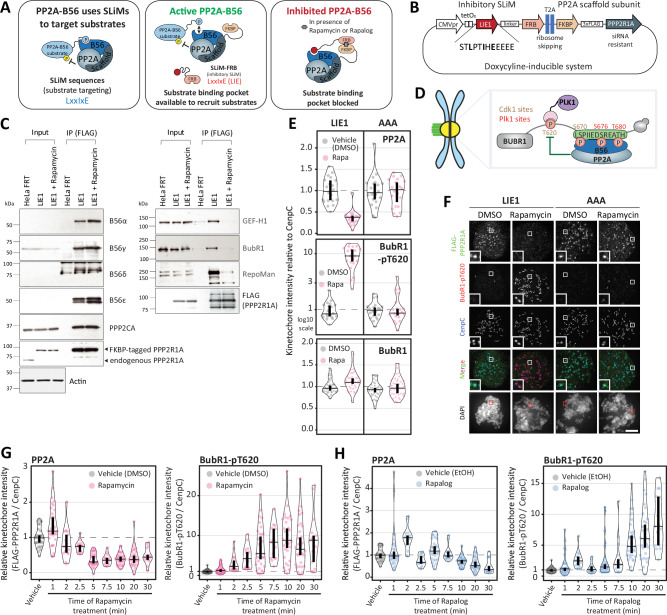
Serine-threonine phosphatases have been challenging to study because of the lack of specific inhibitors. Their catalytic domains are druggable, but these are shared or very similar between individual phosphatase complexes, precluding their specific inhibition. Instead, phosphatase complexes often achieve specificity by interacting with short linear motifs (SLiMs) in substrates or their binding partners. We develop here a chemical-genetic system to rapidly inhibit these interactions within the PP2A-B56 family. Drug-inducible recruitment of ectopic SLiMs ("directSLiMs") is used to rapidly block the SLiM-binding pocket on the B56 regulatory subunit, thereby displacing endogenous interactors and inhibiting PP2A-B56 activity within seconds. We use this system to characterise PP2A-B56 substrates during mitosis and to identify a role for PP2A-B56 in allowing metaphase kinetochores to properly sense tension and maintain microtubule attachments. The directSLiMs approach can be used to inhibit any other phosphatase, enzyme or protein that uses a critical SLiM-binding interface, providing a powerful strategy to inhibit and characterise proteins once considered "undruggable".


Conflict of interest statement

Competing interests: The authors declare no competing interests.

Figures

Fig. 1
Fig. 1. A chemical-genetic system to rapidly inhibit PP2A-B56.
A Schematic illustrating the principle of the drug-inducible recruitment of ectopic SLiMs (directSLiMs) system. B Representation of the plasmid used to implement the directSLiMs strategy in HeLa-FRT cells. C. Immunoblot of the PP2A complex (left column) and PP2A substrates (right column), following FLAG immunoprecipitation from nocodazole-arrested HeLa-FRT cells with/without directSLiMLIE1 expression, -/+ rapamycin for 30 min. Representative of two experiments. D Schematic illustrating how PP2A-B56 regulates the phosphorylation of the T620 site on BUBR1. E–F Quantifications (E) and representative example immunofluorescence images (F) to show the effects of PP2A-B56 inhibition on the levels of FLAG-PPP2R1A, BUBR1-pT620 and BUBR1 at unattached kinetochores in nocodazole-arrested HeLa-FRT cells expressing the directSLiMsLIE1/AAA and treated with vehicle or rapamycin for 20 min. Kinetochore intensities from 20-40 cells, 2–4 experiments. The insets show magnifications of the outlined regions. Scale bars: 5 μm. Inset size: 1.5μm G–H. Effects of PP2A-B56 inhibition on the levels of FLAG-PPP2R1A and BUBR1-pT620 at unattached kinetochores, in nocodazole-arrested HeLa-FRT cells expressing directSLiMLIE1 and treated with rapamycin (G) or rapalog (H) for 0-30 mins. Kinetochore intensities from 15–30 cells, 1–2 experiments. Source data are provided as a Source Data file. Data information: Kinetochore intensities in E–H are normalized to directSLiMLIE1 vehicle condition. Violin plots show the distributions of kinetochore intensities between cells. For each violin plot, each dot represents an individual cell, the horizontal line represents the median and the vertical one the 95% CI of the median, which can be used for statistical comparison of different conditions (see Methods).
Fig. 2
Fig. 2. Global protein phosphorylation changes after PP2A-B56 inhibition in directSLiMLIE1 cells.
A Experimental design: mitotically arrested directSLiMLIE1 and HeLa-FRT cells were treated with rapamycin, rapalog or vehicle before harvest (n = 3 for each condition). Following cell lysis, proteins were digested, and peptides were TMT-labeled. Next, phosphopeptides were enriched with Ti-IMAC magnetic beads, followed by high-pH reversed-phase fractionation and measurement by LC-MS. B Zoomed-in right upper quadrant of volcano plot showing upregulated phosphorylation sites after treatment with rapamycin or rapalog. Solid dots are either validated substrates, predicted substrates or interactors of validated PP2A-B56 substrates (see Methods for details). Only hits above a -Log10(P value = 0.05) (Two- sided Student’s t-test) and >1.5-fold change are color coded. C Heat map of phosphorylation sites upregulated after PP2A-B56 inhibition in directSLiMLIE1 cells. Each row represents the intensity of a phosphorylation site (n = 187). Intensities are normalised to vehicle-treated condition for each cell line to show the relative fold-change upon rapamycin/rapalog addition. Gene names are added for all phospho-sites from validated substrates or interactors of validated substrates. D Percentage of Aurora kinase, PLK1 and CDK consensus motifs in the increasing and non-changing phosphorylation sites. E Sequence logo of non-proline directed upregulated phosphorylation sites normalized against background (all non-changing phosphosites used as background). Source data are provided as a Source Data file.
Fig. 3
Fig. 3. Basal inhibition of PP2A-B56 is observed in directSLiMLIE1 cells.
A Schematic illustrating kinetochore phosphosites on the BUB complex and KNL1 that are regulated by PP2A-B56. BC Effects of PP2A-B56 inhibition on the levels of BUB1-pT609, BUB1-pT461 and BUB1 (B, left) or KNL1-pMELT, KNL1-pRVSF and KNL1 (B, right) or BUBR1-pT620 and BUBR1 (C) at unattached kinetochores, in nocodazole-arrested HeLa-FRT cells with/without directSLiMLIE1 expression and treated with vehicle or rapamycin for 20 min. Kinetochore intensities from 20–30 cells, 2–3 experiments. D Levels of BubR1-pT620 at unattached kinetochores in nocodazole-arrested directSLiMLIE1 cells treated with rapamycin for indicated times. Kinetochore intensities from 30 cells, 3 experiments E Heat map of phosphorylation sites upregulated after PP2A-B56 inhibition in directSLiMLIE1 cells. Each row shows the z-scored intensity of a phosphorylation site (n = 170). Source data are provided as a Source Data file. Data information: Kinetochore intensities in BC are normalized to directSLiMLIE1 vehicle condition. Violin plots show the distributions of kinetochore intensities between cells. For each violin plot, each dot represents an individual cell, the horizontal line represents the median and the vertical one the 95% CI of the median, which can be used for statistical comparison of different conditions (see Methods).
Fig. 4
Fig. 4. Optimization of the directSLiMs strategy to reduce basal inhibition of PP2A-B56.
A Table reporting the tested SLiMs with a lower binding affinity for PP2A-B56 in comparison to LIE1. For each peptide sequence, the SLiM is highlighted in bold. B Effects of PP2A-B56 inhibition on the levels of BUBR1-pT620 at unattached kinetochores (top) and on the mitotic cell fate after nuclear envelope breakdown (NEBD, bottom) in HeLa-FRT cells with/without directSLiMsLIE1-7/AAA expression and treated with vehicle or rapamycin. Top graph: Kinetochore intensities from 20 nocodazole-arrested cells, 2 experiments except HeLa-FRT, DMSO: 10 cells. The treatment with vehicle/rapamycin was performed for 20 min prior fixation. Bottom graph: heatmap showing the mean frequencies of cell fate after NEBD in each condition: 2 experiments, 17–50 cells per condition per experiment (see also Supplementary Fig. 4A). C Table reporting the tested SLiMs with different numbers of acidic residues C-terminal to the LxxIxE motif. The acidic residues restored from LPTI (modified peptide) are highlighted in bold. D Effects of PP2A-B56 inhibition on the levels of BUBR1-pT620 at unattached kinetochores (top) and on the mitotic cell fate after nuclear envelope breakdown (NEBD, bottom) in HeLa-FRT cells with/without directSLiMsLIE1/LIE8-14/AAA expression and treated with vehicle or rapamycin. Top graph: Kinetochore intensities from 20 nocodazole-arrested cells, 2 experiments. The treatment with vehicle/rapamycin was performed for 20 min prior fixation. Bottom graph: heatmap showing the mean frequencies of cell fate after NEBD in each condition: 2 experiments, 42-50 cells per condition per experiment (see also Supplementary Fig. 4B). E Levels of BubR1-pT620, Bub1-pT609, Knl1-pMELT and Knl1-pRVSF at unattached kinetochores, in nocodazole-arrested HeLa-FRT cells expressing directSLiMsLIE1/LIE9 and treated with vehicle for 20 min. Note that the distributions of directSLiMLIE1 and directSliMLIE9 are also shown in Supplementary Fig. 4D. Kinetochore intensities from 20 to 30 cells, 2–3 experiments. Source data are provided as a Source Data file. Data information: Kinetochore intensities in B, D and E are normalized to directSLiMLIE1 vehicle condition. Violin plots show the distributions of kinetochore intensities between cells. For each violin plot, each dot represents an individual cell, the horizontal line represents the median and the vertical one the 95% CI of the median, which can be used for statistical comparison of different conditions (see Methods).
Fig. 5
Fig. 5. Global protein phosphorylation changes after PP2A-inhibition in directSLiMLIE9 cells.
A Experimental design: mitotically arrested directSLiMLIE9 and directSLiMAAA cells were treated with rapamycin, rapalog or vehicle before harvest (n = 3 for each condition). Following cell lysis, proteins were digested, and peptides were TMT-labeled. Next, phosphopeptides were enriched with Fe-NTA spin column, followed by high-pH reversed-phase fractionation and measurement by LC-MS. B Zoomed-in right upper quadrant of volcano plot showing upregulated phosphorylation sites after treatment with rapamycin or rapalog. Solid dots are either validated substrates, predicted substrates or interactors of validated PP2A-B56 substrates (see methods for details). Only hits above a -Log10(P value = 0.05) (Two- sided Student’s t-test) and >1.5-fold change are color coded. C Heat map of phosphorylation sites upregulated after PP2A-B56 inhibition in directSLiMLIE9 cells. Each row represents the intensity of a phosphorylation site (n = 149). Intensities are normalised to vehicle treated condition for each cell line to show the relative fold-change upon rapamycin/rapalog addition. Gene names are added for all phospho-sites from validated substrates or interactors of validates substrates. D Percentage of Aurora kinase, PLK1 and CDK consensus motifs in the increasing and non-changing phosphorylation sites. E Sequence logo of non-proline directed upregulated phosphorylation sites normalized against background (all non-changing phosphosites used as background). F Heat map of phosphorylation sites upregulated after PP2A-B56 inhibition in directSLiMLIE9 cells. Each row represents the z-scored intensity of a phosphorylation site (n = 155). The label (ii) refers to phospho-sites in doubly phosphorylated peptides. Source data are provided as a Source Data file.
Fig. 6
Fig. 6. PP2A-B56 is needed to maintain stable kinetochore-microtubules attachment at metaphase.
A Schematic illustrating known and potential contributions of PP2A-B56 in stabilizing kinetochore-microtubule attachments during mitosis. B Evaluating the effects on chromosome alignment in HeLa-FRT cells with/without directSLiMsLIE9/AAA expression and treated with vehicle or rapamycin. Top panel: protocol used to visualise chromosome alignment in fixed samples (see Methods for details). Bottom panel: graph showing mean frequencies of chromosome misalignments ( ± SEM) of 2 experiments, 100 cells quantified per condition per experiment. Individual data points of the bar charts are provided in Source Data file. C Evaluating the timings of chromosome misalignments during a metaphase arrest, in HeLa HistH1H4C-TagGFP2 cells expressing the directSLiMsLIE1/LIE9/AAA and treated with vehicle or rapamycin. Top panel shows protocol and bottom panel shows timings of chromosome misalignments from 2 experiments, 50 cells quantified per condition per experiment. D Example images of the misalignment timings shown in C. E Kinetochore intensity of NDC80-pS55 in nocodazole-arrested HeLa-FRT cells with directSLiMsLIE1/LIE9/AAA treated with vehicle or rapamycin. 60 cells quantified from 4 repeats. F Evaluating the timings of chromosome misalignments during a metaphase arrest, in HeLa HistH1H4C-TagGFP2 cells expressing the directSLiMLIE1 and treated with/without rapamycin and the AurBi ZM-447439, as indicated. Top panel shows protocol and bottom panel shows timings of chromosome misalignments from 2 experiments, 50 cells quantified per condition per experiment. G Example images of the misalignment timings shown in F. H Fixed chromosome alignment assays. Top panel shows protocol and bottom panel mean mean frequencies of chromosome misalignments ( ± SEM) of 2 experiments, 100 cells quantified per condition per experiment. Individual data points of the bar charts are provided in Source Data file. I Kinetochore intensities of SKAP from cells treated as in H. SKAP is a marker of mature end-kinetochore microtubules attachments. 20 cells quantified from 2 repeats. Source data are provided as a Source Data file. Data information: Violin plots show the distributions of misalignment timings between cells. For each violin plot, each dot represents an individual cell, the horizontal line represents the median and the vertical one the 95% CI of the median, which can be used for statistical comparison of different conditions (see Methods). Scale bar: 10 μm.

References

    1. Brautigan, D. L. & Shenolikar, S. Protein Serine/Threonine Phosphatases: Keys to Unlocking Regulators and Substrates. Annu Rev. Biochem87, 921–964 (2018). - PubMed
    1. Kokot, T. & Köhn, M. Emerging insights into serine/threonine-specific phosphoprotein phosphatase function and selectivity. J. Cell Sci.135, (2022). - PubMed
    1. Nguyen, H. & Kettenbach, A. N. Substrate and phosphorylation site selection by phosphoprotein phosphatases. Trends Biochem Sci.48, 713–725 (2023). - PMC - PubMed
    1. Brautigan, D. L. Protein Ser/Thr phosphatases-the ugly ducklings of cell signalling. Febs j.280, 324–345 (2013). - PubMed
    1. Fowle, H., Zhao, Z. & Graña, X. PP2A holoenzymes, substrate specificity driving cellular functions and deregulation in cancer. Adv. Cancer Res144, 55–93 (2019). - PMC - PubMed

Substances

LinkOut - more resources