. 2025 Apr 22;44(4):115479.
doi: 10.1016/j.celrep.2025.115479. Epub 2025 Mar 29.

Pyroptosis of pulmonary fibroblasts and macrophages through NLRC4 inflammasome leads to acute respiratory failure

Affiliations

Pyroptosis of pulmonary fibroblasts and macrophages through NLRC4 inflammasome leads to acute respiratory failure

Yan Zhang et al. Cell Rep. .

Abstract

The NAIP/NLRC4 inflammasome plays a pivotal role in the defense against bacterial infections, with its in vivo physiological function primarily recognized as driving inflammation in immune cells. Acute lung injury (ALI) is a leading cause of mortality in sepsis. In this study, we identify that the NAIP/NLRC4 inflammasome is highly expressed in both macrophages and pulmonary fibroblasts and that pyroptosis of these cells plays a critical role in lung injury. Mice challenged with gram-negative bacteria or flagellin developed lethal lung injury, characterized by reduced blood oxygen saturation, disrupted lung barrier function, and escalated inflammation. Flagellin-induced lung injury was protected in caspase-1 or GSDMD-deficient mice. These findings enhance our understanding of the NAIP/NLRC4 inflammasome's (patho)physiological function and highlight the significant role of inflammasome activation and pyroptosis in ALI during sepsis.

Keywords: CP: Immunology; inflammasome; lung injury; pyroptosis; sepsis.


Conflict of interest statement

Declaration of interests The authors declare no competing interests.

Figures

Figure 1.
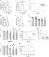
Figure 1.. Flagellin-induced acute lung injury requires NAIP/NLRC4 inflammasome
(A and B) Kaplan-Meier survival plots (A) and monitoring of arterial oxygen saturation (B) were conducted in WT or Casp1−/− mice intravenously injected with 5 μg LFn-flagellin/PA. (C) Lung capillary filtration coefficient (Kf,c) was measured before or 1 h after flagellin administration. (D and E) Kaplan-Meier survival plots for WT mice, TLR5−/−, or NAIP−/−-deficient mice challenged with 5 μg LFn-flagellin/PA (D), and monitoring of arterial oxygen saturation using infrared pulse oximetry (E). (F and G) WT mice intravenously injected with 5 μg LFn-FlicEC/PA or LFn-FlicPA/PA. Arterial oxygen saturation (F) and lung Kf,c were determined (G). (H and I) WT mice injected intravenously with 5 μg LFn-EprJ/PA, with determination of arterial oxygen saturation (H) and lung Kf,c (I). Error bars represent the mean ± SEM. *p < 0.05; **p < 0.01;****p < 0.0001; ns, not significant, by log rank (Mantel-Cox) test (A and D), two-tailed unpaired t test (I), one-way ANOVA (G and H), or two-way ANOVA with Holm-Sidak multiple comparisons test (B, C, E, and F).
Figure 2.
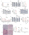
Figure 2.. Lung cell pyroptosis plays a critical role in inflammasome-induced lung injury
(A–C) WT mice received intravenous injections of PA (Ctrl), LFn-flagellin plus PA (3 μg of each per mouse), or the same dose of LFn-flagellin-3A mutant plus PA. Blood was collected 90 min post-injection. PT (A) and TAT (B) were measured. Kaplan-Meier survival plot is shown (C). (D–F) WT mice were injected intravenously with a rat immunoglobulin G or a rat anti-mouse TF-neutralizing antibody 1H1 (8 mg/kg). After 2 h, mice received flagellin, and blood was collected 90 min later. Prothrombin time (D), plasma TAT concentrations (E), and arterial oxygen saturation (F) were measured. (G–I) WT mice or GSDMD−/− mice were injected intravenously with 5 μg LFn-flagellin/PA. Arterial oxygen saturation (G) and Kaplan-Meier survival plots are shown (H), and lung Kf,c was determined (I). (J and K) WT mice received intravenously IL-1RA (1 mg/kg body weight) 10 min prior to injection of LFn-flagellin/PA. Arterial oxygen saturation and Kaplan-Meier survival plots are shown. Error bars represent the mean ± SEM. **p < 0.01;****p < 0.0001; ns, not significant, by log rank (Mantel-Cox) test (C, H, and K), two-tailed unpaired t test (D, E, and I), one-way ANOVA (A and B), or two-way ANOVA with Holm-Sidak multiple comparisons test (F, G, and J).
Figure 3.
Figure 3.. Flagellin-induced lung injury extends beyond macrophage pyroptosis
(A and B) WT or TLR5−/− mice were intravenously injected with 5 μg LFn-flagellin/PA. After 1 h, the total number of macrophages (A) and cytokine levels (B) in BAL fluid were measured. (C) WT mice received PBS, control liposomes (Lipo), or clodronate-containing liposomes (Cldn) 24 h prior to injection of 5 μg LFn-flagellin/PA. Arterial oxygen saturation was monitored. (D and E) Lung cells were incubated with PA (Ctrl) or 1 μg/mL LFn-flagellin/PA for 6 h. LDH concentration (D) and levels of p20 caspase-1 and p17 IL-1β by immunoblotting (E) were detected. (F) Lung cells were pre-incubated with PBS, 100 μg/mL Lipo, or Cldn, then transfected with PA (Ctrl) or 1 μg/mL LFn-flagellin/PA for 6 h. LDH concentrations were measured. (G and H) Epithelial (G) and endothelial (H) cells were incubated with PA (Ctrl) or 1 μg/mL LFn-flagellin/PA for 6 h. As a positive control, cells were primed with 1 μg/mL LPS for 4 h, followed by stimulation with 20 μM nigericin for 4 h, with or without caspase-1 inhibitor Ac-YVAD-cmk (5 μM). LDH concentrations in the supernatant were measured to determine cytotoxicity. Error bars represent the mean ± SEM. *p < 0.05; **p < 0.01; ***p < 0.001; ****p < 0.0001; ns, not significant, by two-tailed unpaired t test (A and D), one-way ANOVA (G and H), or two-way ANOVA with Holm-Sidak multiple comparisons test (B, C, and F).
Figure 4.
Figure 4.. Pulmonary fibroblast is responsible for flagellin-induced lung injury
(A) The procedure of scRNA-seq and uniform manifold approximation and projection plot displaying distinct changes in different cell types within the lungs following challenge with LFn-flagellin/PA. (B) Fibroblasts isolated from WT mice and Casp1−/− mice were incubated with PA (Ctrl) or 1 μg/mL LFn-flagellin/PA for 6 h. Cytotoxicity was determined by LDH assay. (C) Bone marrow-derived macrophages, as well as lung epithelial, endothelial, and fibroblasts isolated from untreated WT mice were incubated with PA (Ctrl) or 1 μg/mL LFn-flagellin/PA for 6 h. P20 caspase-1 and p17 IL-1β were detected by immunoblot. (D and E) Significant changes in gene expression in the different cell populations (D), including lipofibroblast (E) population following flagellin treatment. (F) NLRC4 inflammasome components were detected in fibroblasts from lung, heart, and colon tissues of mice. (G) GSDMDfl/fl/Col1a2Cre or GSDMDfl/fl/Col1a2Cre+ mice were injected intravenously with 5 μg LFn-flagellin/PA. Cre+ mice were further treated with Cldn 24 h prior to flagellin injection. Arterial oxygen saturation was monitored using infrared pulse oximetry. Error bars represent the mean ± SEM. *p < 0.05; ****p < 0.0001; ns, not significant, by two-way ANOVA with Holm-Sidak multiple comparisons test (B and G).
Figure 5.
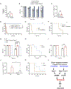
Figure 5.. Caspase-8/GSDME pathway in macrophage contributed to acute lung injury induced by high-dose flagellin
(A and B) WT mice, Casp1−/−, NAIP−/−, or Nlrc4−/− mice were injected with 10 μg LFn-flagellin/PA. Kaplan-Meier survival plots (A) and arterial oxygen saturation were monitored (B). (C) Casp1−/− mice received 10 μg LFn-flagellin/PA. The lung Kf,c was determined. (D) WT mice or TLR5−/−-deficient mice received 10 μg LFn-flagellin/PA. Arterial oxygen saturation was monitored. (E) Total lung cells isolated from Casp1−/− mice were incubated with PA (Ctrl) or 10 μg/mL LFn-flagellin/PA for 6 h. Cytotoxicity was determined by LDH assay. (F) Casp1−/− mice were pre-treated with PBS, Lipo, or Cldn 24 h prior to injection of 10 μg LFn-flagellin/PA. Arterial oxygen saturation was monitored. (G) Total lung cells isolated from WT mice or Casp1−/− mice were pre-incubated with PBS and 100 μg/mL Lipo or Cldn, and then incubated with PA or 1 μg/mL LFn-flagellin/PA for 6 h. LDH in the supernatant was measured. (H) WT, GSDMD−/−, and GSDMD−/−/GSDME−/− mice were injected 10 μg LFn-flagellin/PA. Arterial oxygen saturation was monitored. (I) Total lung cells isolated from WT, GSDMD−/−, and GSDMD−/−/GSDME−/− mice were incubated with PA or 1 μg/mL LFn-flagellin/PA for 6 h. LDH in supernatant was measured. (J and K) WT and GSDMD−/−/GSDME−/− mice were injected 10 μg LFn-flagellin/PA. Histological evaluation of lung morphology (J) and Kaplan-Meier survival plots (K) were measured. Black arrow points to mononuclear cell infiltration in lungs, blue arrow points to thickened alveolar wall, and red arrow points to hemorrhage. Scale bars, 200 μm. Error bars represent the mean ± SEM. *p < 0.05; **p < 0.01; ***p < 0.001; ****p < 0.0001; ns, not significant, by log rank (Mantel-Cox) test (A and K), two-tailed unpaired t test (C and E), or two-way ANOVA with Holm-Sidak multiple comparisons test (B, D, and F–I).
Figure 6.
Figure 6.. Caspase-8-dependent pyroptosis of macrophages contributes to lung injury in the absence of caspase-1
(A) Total lung cells isolated from WT and Casp1−/−/Casp8−/−/Ripk3−/− mice were incubated with 1 μg/mL LFn-flagellin/PA for 6 h. Cell culture supernatants were used to measure LDH concentration. (B–D) Ripk3−/−mice, Casp8−/−/Ripk3−/− mice, and Casp1−/−/Casp8−/−/Ripk3−/− mice received 10 μg LFn-flagellin/PA. Arterial oxygen saturation (B), lung Kf,c (C), and Kaplan-Meier survival plots (D) for flagellin-challenged mice are shown. (E and F) WT (E) and Casp1−/− mice (F) underwent mechanical ventilation and were injected intravenously with 10 μg LFn-flagellin/PA for up to 6 h. Kaplan-Meier survival plots for mice challenged with flagellin are shown. (G) WT, Nlrc4−/−, and Casp1−/−/Casp8−/−/Ripk3−/− mice were injected intraperitoneally with 2 × 10⁸ CFU Salmonella typhimurium for 6 h. Arterial oxygen saturation was measured. (H) WT and Nlrc4−/− mice were injected intraperitoneally with 2 × 10⁸ CFU S. typhimurium. Kaplan-Meier survival plots are shown. (I–K) WT and Nlrc4−/− mice were subjected to the FIP model. Arterial oxygen saturation (I), lung Kf,c (J), and Kaplan-Meier survival analysis (K) following administration of a 40-mg/mL feces solution are shown. (L) Model of lung injury triggered by caspase-1-dependent and -independent inflammasome activation. Error bars represent the mean ± SEM. *p < 0.05; **p < 0.01; ***p < 0.01; ****p < 0.0001; ns, not significant, by log rank (Mantel-Cox) test (D–F, H, and K), two-tailed unpaired t test (A and C), or two-way ANOVA with Holm-Sidak multiple comparisons test (B, G, I, and J).

References

    1. Sundaram B, and Kanneganti TD (2021). Advances in Understanding Activation and Function of the NLRC4 Inflammasome. Int. J. Mol. Sci 22, 1048. - PMC - PubMed
    1. Wei Y, Yang L, Pandeya A, Cui J, Zhang Y, and Li Z (2022). Pyroptosis-Induced Inflammation and Tissue Damage. J. Mol. Biol 434, 167301. 10.1016/j.jmb.2021.167301. - DOI - PMC - PubMed
    1. Miao EA, Alpuche-Aranda CM, Dors M, Clark AE, Bader MW, Miller SI, and Aderem A (2006). Cytoplasmic flagellin activates caspase-1 and secretion of interleukin 1beta via Ipaf. Nat. Immunol 7, 569–575. - PubMed
    1. Miao EA, Mao DP, Yudkovsky N, Bonneau R, Lorang CG, Warren SE, Leaf IA, and Aderem A (2010). Innate immune detection of the type III secretion apparatus through the NLRC4 inflammasome. Proc. Natl. Acad. Sci. USA 107, 3076–3080. - PMC - PubMed
    1. Zhao Y, Yang J, Shi J, Gong YN, Lu Q, Xu H, Liu L, and Shao F (2011). The NLRC4 inflammasome receptors for bacterial flagellin and type III secretion apparatus. Nature 477, 596–600. - PubMed

MeSH terms

LinkOut - more resources