Review
. 2025 Aug 4;27(8):euaf076.
doi: 10.1093/europace/euaf076.

Practical compendium of antiarrhythmic drugs: a clinical consensus statement of the European Heart Rhythm Association of the European Society of Cardiology

Affiliations
Review

Practical compendium of antiarrhythmic drugs: a clinical consensus statement of the European Heart Rhythm Association of the European Society of Cardiology

Jose L Merino et al. Europace. .

Abstract

The European Heart Rhythm Association Practical Compendium of Anti-arrhythmic Drugs (AADs) offers advice on these drugs, focusing on their clinical use and the global impact of cardiac arrhythmias. This document aims to provide practical instructions to clinicians in arrhythmia management through pharmacological strategies. The compendium highlights persistent challenges in arrhythmia treatment, including clinical constraints, procedural risks, and the complexity of certain arrhythmias. Notably, atrial fibrillation is highly prevalent, and the demand for invasive treatment often surpasses the capacity of existing healthcare systems. As a result, pharmacological management remains essential. This is particularly relevant for patients with cardiac implantable electronic devices or channelopathies, where ablation is often not a suitable option. Anti-arrhythmic drugs play a pivotal role in these scenarios. The compendium introduces the ABC framework for AAD therapy: A (Appropriate therapy), for patients in whom AADs are the best therapeutic option; B (Backup therapy), as adjunctive treatment to invasive procedures, such as catheter ablation; and C (Complementary therapy), in combination with other therapies. The document provides detailed insights into the mechanisms of action, efficacy, safety profiles, and drug interactions of each class of AADs. Additionally, the compendium covers practical considerations, including initiation, combination strategies, monitoring, follow-up, special populations, and adverse effect management, with an emphasis on pro-arrhythmia risk mitigation. It also explores the integration of AADs with other therapeutic modalities, promoting a synergistic approach to optimize patient outcomes. In summary, this compendium serves as an indispensable resource for clinicians, offering practical advice and evidence-based insights to navigate the complexities of arrhythmia management effectively.

Keywords: Adverse drug reactions; Anti-arrhythmic drug combinations; Anti-arrhythmic drugs; Arrhythmia; Atrial fibrillation; Drug interactions; Mechanisms; Pharmacology; Ventricular arrhythmias.


Conflict of interest statement

Conflict of interest: A.G. has received funding from the EU Horizon 2020 programme: MAESTRIA Consortium grant number 952166 and speaker fees from Astra Zeneca, Boehringer Ingelheim, BMS/Pfizer, Daiichi Sankyo, and Medtronic. A.J.C. has received personal consulting fees from Acesion, InCarda, Menarini, Milestone, Sanofi, Anthos, Bayer, Daiichi Sankyo, Pfizer, Abbott, Biosense Webster, Biotronik, Boston Scientific, Medtronic, GlaxoSmithKline, and Johnson & Johnson. C.B.-L. has received fees and honoraria for lectures, education, and scientific advice from Abbott, Biosense Webster, Bayer, Sanofi, Organon, Philips, Medtronic, Boston Sci, and Cathprint. D.D. has received fees and honoraria for lectures and education from Daiichi Sankyo. G.B. reports small speaker fees from Bayer, Boehringer Ingelheim, Boston, Daiichi Sankyo, Janssen, and Sanofi outside of the submitted work. He is also the principal investigator of the ARISTOTELES (Applying ARtificial Intelligence to define clinical trajectorieS for personalized predicTiOn and early deTEction of comorbidity and muLtimorbidity pattErnS) project that received funding from the European Union within the Horizon 2020 research and innovation programme (grant no. 101080189). H.J.G.M.C. has received fees and honoraria for lectures, education, and scientific advice from InCarda Therapeutics, Roche, Sanofi, Atricure, Medtronic, and Armgo. J.L.M. has received fees and honoraria for lectures, education, and scientific advice from Abbott, Biosense Webster, Biotronik, iRhythm Technologies, MicroPort, and Zoll. He is also a member of the steering committe in the EHRA-PATHS (Addressing Multimorbidity in Elderly Atrial Fibrillation Patients Through Interdisciplinary, Tailored, Patient-Centered Care Pathways, GA 945260) and PROFID (Implementation of Personalized Risk Prediction and Prevention of Sudden Cardiac Death After Myocardial Infarction, GA 847999) projects, both funded by the European Union under the Horizon 2020 Research and Innovation Programme. J.A.R. reports being an investigator for Sanofi, InCarda Therapeutics, Johnson & Johnson, and Amarin and as a consultant for Sanofi and Acesion. J.T.-F. has received fees and honoraria for lectures, education, and scientific advice from Cytokinetics, Johnson and Johnson, MicroPort, Leo Pharma, and Boston Scientific. SHH has received fees and honoraria for lectures, education, and scientific advice from Sanofi, BI, Pfizer, BMS, Daiichi, and Incardia. The remaining authors declared no conflict of interest.

Figures

Graphical Abstract
Graphical Abstract
Figure 1
Figure 1
Schematic representation of the three main states (resting, activated, and inactivated) of an ionic channel in the cellular surface membrane of a cardiomyocyte. During the resting phase (left panel), the influx of ions into the cell is not possible (short arrow) because the channel remains closed (horizontal rectangles). Once the channel is activated (central panel), ions can enter the cell (long arrow) through the open channel (small oblique rectangles). Following activation, the channel transitions to an inactivated state (right panel, inferior horizontal rectangle), preventing further ion influx. Different anti-arrhythmic drugs (AADs) (e.g. flecainide) exhibit specific affinity and preferentially bind to particular states of the channel (e.g. the activated state).
Figure 2
Figure 2
Schematic representation of the effects of flecainide (A and B) and sotalol (C) on the transmembrane action potential during sinus rhythm (SR) (A), atrial fibrillation (AF) (B), and sinus bradycardia (C). The figure also illustrates their potential anti-arrhythmic and pro-arrhythmic effects on AF (ECG in panel B) and sinus bradycardia (ECG in panel C), respectively. Flecainide (green polygon) binds to the sodium channel (Na+ Ch) primarily in its activated (slightly separated red rectangles) and inactivated (closely aligned grey rectangles) states. Its maximal effect is observed during tachycardia, as the shortened action potential duration keeps the sodium channel in these states more frequently. This use-dependent property enables flecainide to effectively block the activation front, contributing to the termination of atrial fibrillation (AF). Additionally, its very slow dissociation kinetics and strong binding to the inactivated state play a crucial role in prolonging post-repolarization refractoriness—a key mechanism underlying its anti-arrhythmic efficacy but also a potential contributor to pro-arrhythmia. In contrast, sotalol (red polygon) binds to several potassium channels (K+Ch) mostly during its resting state (closely aligned blue rectangles). Its maximum effect occurs in bradycardia, where the channel remains in this state for a longer duration. This reverse use-dependent effect leads to prolonged action potential duration and QT interval prolongation, which can trigger early afterdepolarizations (EADs) and ventricular tachycardia, including torsades de pointes (TdP). Downward curved arrows represent anti-arrhythmic drug (AAD) binding to the ion channel, while upward curved arrows indicate the absence of binding.
Figure 3
Figure 3
Schematic representation of intestinal absorption, tissue storage, and hepatic and renal excretion pathways for commonly affected anti-arrhythmic drugs (AADs). Box A: Intestinal absorption occurs through epithelial cells (enterocytes) of the small intestine. However, P-glycoprotein (P-gp) in enterocytes actively effluxes a portion of certain drugs back into the intestinal lumen, reducing systemic absorption. Box B: Lipophilic drugs tend to accumulate in fat-rich tissues and organs, such as the lungs, liver, thyroid, and adipose tissue (primary tissues of accumulation listed in brackets). Box C: Hydrophilic drugs exhibit minimal or no tissue accumulation and distribute predominantly in the extracellular fluid,—except for digoxin, which primarily accumulates in cardiac muscle. Box D: Drugs are metabolized by the liver and excreted via bile into faeces. Box E: Renal clearance eliminates drugs or their metabolites through the kidneys. Approximate percentages of drug efflux and elimination are indicated in the respective boxes. Flecainide is partially eliminated by the kidneys (~30–40%), and impaired renal function can lead to drug accumulation and increased risk of proarrhythmia. CCB, calcium channel blocker.
Figure 4
Figure 4
Twelve-lead electrocardiograms (ECGs) illustrating the effect of quinidine on the QT interval in a female patient with no structural heart disease and a history of atrial fibrillation. (A) Baseline ECG recorded prior to quinidine administration, showing a normal QTc interval duration (two-arrowhead line). (B) ECG following quinidine administration, revealing marked QT interval prolongation, indicative of its effect on ventricular repolarization. This underscores the potential for pro-arrhythmic effects, even in the absence of structural heart disease.
Figure 5
Figure 5
Electrocardiogram (ECG) tracings of Leads II and III illustrating the termination of ventricular tachycardia (VT) after a 15 min infusion of procainamide in a patient with structural heart disease (old myocardial infarction). The tracings show VT transitioning to sinus rhythm after procainamide administration, demonstrating its anti-arrhythmic efficacy in managing VT in the presence of underlying myocardial scarring. Electrocardiogram was recorded at a speed of 25 mm/s and a sensitivity of 10 mm/mV.
Figure 6
Figure 6
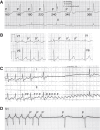
Single-lead (A) and 12-lead electrocardiogram (ECG) tracings (B and C) demonstrating the progression of atrial arrhythmias in a 57-year-old hypertensive male patient taking 200 mg/day of flecainide for paroxysmal atrial fibrillation (AF). (A) Baseline ECG showing AF at presentation. (B) After a few days of flecainide therapy, the patient developed atrial flutter (AFL) with 1:1 atrioventricular (AV) conduction, non-specific intraventricular conduction disturbance, and rapid ventricular response with RR intervals of 280 ms, mimicking ventricular tachycardia (VT). (C) Following the administration of 5 mg of i.v. atenolol, the AV conduction ratio changed to 3:1. This resulted in the resolution of LBBB and narrowing of the QRS complex, making the flutter waves apparent, with a cycle length of 240 ms (cycle length shortened by 40 ms after a partial washout effect of flecainide). This case illustrates flecainide-induced pro-arrhythmia with AFL, and the diagnostic clarity achieved through rate control and conduction ratio alteration. The electrocardiogram was recorded at a speed of 25 mm/s and a sensitivity of 10 mm/mV.
Figure 7
Figure 7
Electrocardiogram (ECG) tracing of Lead II demonstrating the termination of atrial fibrillation (AF) after a 7 min infusion of 350 mg of vernakalant in a patient with no structural heart disease. The transition to sinus rhythm is marked by the appearance of normal P waves (P), indicating successful restoration of organized atrial activity. This highlights the efficacy of vernakalant in achieving cardioversion in patients with AF. Electrocardiogram was recorded at a speed of 5 mm/s and a sensitivity of 10 mm/mV.
Figure 8
Figure 8
Electrocardiogram (ECG) tracings illustrating the effects of adenosine on different atrial rhythms: sinus rhythm (A), atrial tachycardia (B), atrial flutter (AFL, C), and paroxysmal supraventricular tachycardia (PSVT, D). (A) Sinus rhythm at a rate of 88 b.p.m. slows significantly with PR interval prolongation following adenosine infusion. (B) Atrial tachycardia at 125 b.p.m., characterized by a non-sinus P-wave morphology (P′). Initially, conduction is 1:1 atrioventricular (AV) (left). After adenosine administration, conduction changes to 2:1 AV (right) without a significant change in atrial rate. (C) Common AFL with 2:1 AV conduction. Adenosine-induced AV block reveals prominent F waves, enhancing visualization of the flutter waves. (D) Termination of atrioventricular re-entrant tachycardia (AVRT) mediated by a left-sided concealed accessory pathway. Termination occurs after AV node conduction block, interrupting conduction of the final retrograde P′ wave and restoring sinus rhythm. All ECGs were recorded at a speed of 25 mm/s and a sensitivity of 10 mm/mV.
Figure 9
Figure 9
Twelve-lead electrocardiograms (ECGs) of a patient with Wolff–Parkinson–White (WPW) syndrome caused by a right posteroseptal accessory pathway which becomes more prominent following adenosine infusion. (A) Pre-excitation becomes more apparent following the infusion of 12 mg of adenosine, which blocks conduction through the atrioventricular (AV) node. This is evidenced by a pronounced delta wave, indicative of increased conduction via the accessory pathway. (B) Pre-excitation diminishes as AV nodal conduction resumes after the effects of adenosine dissipate, reducing the contribution of the accessory pathway to ventricular depolarization. These findings demonstrate the dynamic inter-play between AV nodal conduction and accessory pathway activation in WPW syndrome, highlighting the diagnostic utility of adenosine in unmasking pre-excitation. The recordings were obtained at a speed of 25 mm/s and a sensitivity of 10 mm/mV.
Figure 10
Figure 10
Anti-arrhythmic drug (AAD) selection based on cardiac substrate and main target of action. This figure advises on the selection of AADs based on their primary target [sinus/AV nodes vs. working atrial (A) and ventricular (V) myocardium] and the presence of ventricular dysfunction, scarring, or heart failure (HF). Class 0, II, and IV agents [e.g. ivabradine, β-blocker (BB), Ca2+ channel blocker (CCB), and digoxin] are preferred for rate control by acting on the sinus and AV nodes. Class I and III agents (e.g. flecainide, propafenone, amiodarone, dronedarone, sotalol, and dofetilide) are used for rhythm control, but their choice depends on the structural integrity of the ventricles. Structural heart disease discourages Class I use, favouring Class III instead. In HF, amiodarone is the preferred option, while other AADs are generally avoided to prevent worsening of the condition. aIvabradine is primarily advised for slowing the sinus rate, with some evidence suggesting it may also influence AV nodal conduction. bBBs and CCBs also affect cardiac tissues beyond the sinus and AV nodes and may be the AADs of choice for certain disorders, such as ectopic atrial tachycardia (AT) or idiopathic fascicular ventricular tachycardia (VT), respectively. cDigoxin is less effective in sinus tachycardia compared with BBs or CCBs. However, digoxin toxicity can lead to severe bradycardia, sinus arrest, or junctional escape rhythms due to excessive vagal stimulation. dClass I and III agents also influence the sinus node and AV conduction but are not the preferred choices for this purpose. eDronedarone is not advised in patients with symptomatic HF or left ventricular ejection fraction (LVEF) <40%. fDofetilide does not worsen survival in HF with reduced ejection fraction (HFrEF) but can prolong the QT interval and cause torsades de pointes. gSotalol is not advised in patients with advanced HF or severe left ventricular dysfunction (LVEF <35%) due to the risk of worsening HF. VW, Vaughan Williams AAD classification.
Figure 11
Figure 11
Schematic representation of the preferred anti-arrhythmic drugs (AADs) for prevention of atrial arrhythmias. The figure serves as a general reference for selecting the most appropriate drug; however, the final choice—or consideration of alternative therapeutic options [e.g. catheter ablation for cavotricuspid isthmus–dependent atrial flutter (AFL)]—is based on the general patient characteristics and conditions, as outlined in the relevant sections of this document. Additionally, not all AADs are available in all regions. For secondary or alternative drug options, refer to Table 3. AAs, atrial arrhythmias; AF, atrial fibrillation; LVEF, left ventricular ejection fraction; SHD, structural heart disease.
Figure 12
Figure 12
Schematic representation of the advised anti-arrhythmic drugs (AADs) for prevention of monomorphic ventricular arrhythmias. The figure serves as a general reference for selecting the most appropriate drug; however, the final choice—or consideration of alternative therapeutic options [e.g. catheter ablation for idiopathic right ventricular outflow tract (RVOT) premature ventricular contractions (PVCs)]—is advised to be based on the general patient characteristics and conditions, as outlined in the relevant sections of this document. Additionally, not all AADs are available in all regions. For secondary or alternative drug options, refer to Table 3. Adrenergic PVCs/VT are characterized by an increased burden and/or severity in response to exercise or mental stress. Ca2, calcium; LVEF, left ventricular ejection fraction; MVT, monomorphic VT; SHD, structural heart disease; VT, ventricular tachycardia.
Figure 13
Figure 13
Twelve-lead electrocardiogram (ECG) demonstrating QT interval prolongation and a 5.5 s run of non-sustained polymorphic ventricular tachycardia [torsade de pointes (TdP)] following a post-extrasystolic pause in a patient with hypomagnesaemia. This highlights the association between electrolyte imbalances, prolonged repolarization, and pro-arrhythmic events such as TdP. The typical TdP twisting pattern of QRS complexes around the isoelectric line (horizontal line) is marked with arrows of varying amplitude above lead V1. Cycle lengths and QT intervals are annotated with black and red numbers, respectively. Torsade de pointes is triggered by a pause (two-arrowhead line) that further prolongs the QT interval. The ECG was recorded at a speed of 25 mm/s and a sensitivity of 10 mm/mV.
Figure 14
Figure 14
Schematic representation of the advised anti-arrhythmic drugs (AADs) for termination of atrial fibrillation (AF). The figure serves as a general reference for selecting the most appropriate drug; however, the final choice is advised to be done by patient-specific characteristics and conditions, as detailed in the various sections of this document. Additionally, drug availability plays a crucial role in decision-making. For example, vernakalant is available in many European countries but not in the United States. Flecainide and propafenone i.v. are not available in many American countries. Antazoline is primarily produced and marketed in Poland, where it is registered for anti-arrhythmic use, though its availability in other European countries remains limited. Pilsicainide is primarily approved and used in Japan and Korea, but its presence in Europe is scarce. Cibenzoline is also used in Japan and has been available in certain European countries. aClass Ic AADs are contraindicated in patients with SHD, HF, or significant conduction disturbances due to the risk of pro-arrhythmia and conduction block. bOral ranolazine has been used off-label for AF conversion in patients with ischaemic heart disease. However, it is advised to be used with caution in NYHA Class III andIV HF and avoided in patients with QT prolongation due to the risk of pro-arrhythmia. AF, atrial fibrillation; HF, heart failure; NYHA, New York Heart Association functional class; i.v., intravenous; p.o., per os; SHD, structural heart disease.
Figure 15
Figure 15
Twelve-lead electrocardiogram (ECG) illustrating the termination of haemodynamically tolerated monomorphic ventricular tachycardia (VT) following the infusion of amiodarone in an 81-year-old female patient with a history of anterior wall myocardial infarction. (A) Initial VT with a cycle length of 330 ms during i.v. amiodarone infusion, showing no significant change in cycle length. (B) Subsequent change in VT morphology and acceleration to a cycle length of 240 ms, followed by VT termination and resumption of sinus rhythm. This case highlights the dynamic response of VT to anti-arrhythmic therapy. The ECG was recorded at a speed of 25 mm/s and a sensitivity of 10 mm/mV.
Figure 16
Figure 16
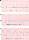
Electrocardiogram (ECG) tracings (Lead III and monitor lead) illustrating the progression of atrial fibrillation (AF) and its response to 300 mg/day flecainide, culminating in ventricular tachycardia (VT) in a 66-year-old female with no underlying heart disease. (A) Baseline AF at a heart rate of 105 b.p.m. with a QRS duration of 90 ms. (B) After administration of 300 mg of flecainide, QRS duration prolongs to 120 ms, while the heart rate remains unchanged at 105 b.p.m. (C) Spontaneous acceleration of AF to 180 b.p.m. leads to further QRS widening to 210 ms, and regularization (atrial flutter conversion with bundle-branch blockvs VT) attributed to the use-dependent effect of flecainide. This sequence underscores the potential pro-arrhythmic effects of flecainide in AF management, if a high dosage is used. The ECG was recorded at a speed of 25 mm/s and a sensitivity of 10 mm/mV.
Figure 17
Figure 17
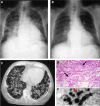
Chest X-ray (A and B), computed tomography (CT) scan (C), and microscopic views of a lung biopsy (D and E) from a 77-year-old former smoker with a history of old myocardial infarction and preserved left ventricular ejection fraction, taking 200 mg/day of amiodarone for paroxysmal atrial fibrillation. The patient presented with cough, dyspnoea, and weight loss. Findings were suggestive of amiodarone-induced lung toxicity. (A) Chest X-ray at presentation showing a diffuse alveolar-interstitial pattern indicative of pulmonary involvement. (B) Follow-up chest X-ray after 3 months of amiodarone withdrawal and steroid therapy showing resolution of lung abnormalities. (C) Computed tomography scan confirming the diffuse alveolar-interstitial pattern at the initial presentation. (D) Optical microscopy (haematoxylin–eosin stain) of lung parenchyma showing clusters of alveolar macrophages (arrows) with foam-like cytoplasmic changes, characteristic of amiodarone toxicity. (E) Electron microscopy of the biopsy sample revealing phospholipid inclusions (red arrow) in macrophages, further confirming the diagnosis of amiodarone-induced pulmonary toxicity. This case underscores the potential for severe pulmonary adverse effects associated with amiodarone therapy and the potential reversibility of findings following drug discontinuation and appropriate treatment.
Figure 18
Figure 18
Single-lead and 12-lead electrocardiograms (ECGs) of a 14-year-old girl with no prior heart disease shortly after ingesting 24 flecainide tablets (2400 mg) in a suicide attempt. (A) The ECG recorded shortly after ingestion shows severe sinus bradycardia with low-amplitude P waves and an extremely wide QRS complex, indicative of significant sodium channel blockade caused by flecainide toxicity. (B) Following resuscitation efforts with sodium chloride, bicarbonate, isoproterenol, and dopamine/dobutamine, the ECG shows sinus tachycardia with less pronounced QRS broadening and repolarization changes. (C) After 9 h of treatment, the ECG demonstrates a decrease of QRS duration and resolution of repolarization abnormalities. Non-captured ventricular temporary pacemaker spikes are also visible. These findings illustrate the severe cardiotoxic effects of flecainide overdose, the dynamic ECG changes during treatment, and the efficacy of intensive medical intervention in reversing these abnormalities. The ECGs were recorded at a speed of 25 mm/s and a sensitivity of 10 mm/mV.
Figure 19
Figure 19
Twelve-lead electrocardiograms (ECGs) of a 22-year-old woman with no prior heart disease presenting with ventricular fibrillation and pleomorphic ventricular arrhythmias. The patient was initially treated with oral quinidine and was later diagnosed with Type II long QT syndrome. The ECG demonstrates inverted T waves (T) across several leads and an extremely prolonged QT interval. This case underscores the pro-arrhythmic potential of quinidine in patients with underlying repolarization disorders. The recordings were obtained at a speed of 25 mm/s and a sensitivity of 10 mm/mV.
Figure 20
Figure 20
Electrocardiogram (ECG) tracings of leads V1–V6 (A) and V1–V3 (B) illustrating the dynamic changes in two patients with Brugada syndrome at baseline, during isoproterenol infusion, and following i.v. administration of flecainide (2 mg/kg). (A) Baseline ECG shows mild ST-segment elevation (1.5 mm) in V2, measured at 80 ms from the J point. Following flecainide infusion, ST-segment elevation increases significantly to 4.5 mm. (B) In another patient, baseline ECG reveals ST elevation and T-wave inversion in V2. These abnormalities normalize during isoproterenol infusion but are markedly exaggerated following flecainide administration. The ECG was recorded at a speed of 25 mm/s and a sensitivity of 10 mm/mV.
Figure 21
Figure 21
Schematic representation of the advised first-line anti-arrhythmic drugs (AADs) for prevention of polymorphic ventricular arrhythmias. The figure provides a general reference for selecting the most appropriate drug; however, the final choice is advised to be based on additional patient characteristics and conditions, as detailed in the various sections of this document. BrS, Brugada syndrome; CPVT, catecholaminergic polymorphic ventricular tachycardia; ERS, early repolarization syndrome; LQTS, long QT syndrome; PVT, polymorphic ventricular tachycardia; SQTS, short QT syndrome.

References

    1. Saksena S, Ken-Opurum J, McKindley DS, Preblick R, Rashkin J, Aldaas OM et al. Arrhythmia recurrence and rhythm control strategies after catheter ablation of newly diagnosed atrial fibrillation (ARRC-AF study). JACC Clin Electrophysiol 2025:S2405-500X(24)01027-2. - PubMed
    1. Heijman J, Voigt N, Nattel S, Dobrev D. Cellular and molecular electrophysiology of atrial fibrillation initiation, maintenance, and progression. Circ Res 2014;114:1483–99. - PubMed
    1. Landstrom AP, Dobrev D, Wehrens XHT. Calcium signaling and cardiac arrhythmias. Circ Res 2017;120:1969–93. - PMC - PubMed
    1. Nattel S, Heijman J, Zhou L, Dobrev D. Molecular basis of atrial fibrillation pathophysiology and therapy: a translational perspective. Circ Res 2020;127:51–72. - PMC - PubMed
    1. Heijman J, Ghezelbash S, Dobrev D. Investigational antiarrhythmic agents: promising drugs in early clinical development. Expert Opin Investig Drugs 2017;26:897–907. - PMC - PubMed

Substances