. 2025 Apr;27(4):e70018.
doi: 10.1002/jgm.70018.

Adoptive Transfer of T Cells as a Potential Therapeutic Approach in the Bleomycin-Injured Mouse Lung

Affiliations

Adoptive Transfer of T Cells as a Potential Therapeutic Approach in the Bleomycin-Injured Mouse Lung

Seyran Mutlu et al. J Gene Med. 2025 Apr.

Abstract

Background: Idiopathic pulmonary fibrosis (IPF) is a lethal disease with an unknown etiology and complex pathophysiology that are not fully understood. The disease involves intricate cellular interplay, particularly among various immune cells. Currently, there is no treatment capable of reversing the fibrotic process or aiding lung regeneration. Hepatocyte growth factor (HGF) has demonstrated antifibrotic properties, whereas the adoptive transfer of modified T cells is a well-established treatment for various malignancies. We aimed to understand the dynamics of T cells in the progression of lung fibrosis and to study the therapeutic benefit of adoptive T cell transfer in a bleomycin-injured mouse lung (BLM) model.

Methods: T cells were isolated from the spleen of naïve mice and transfected in vitro with mouse HGF plasmid and were administered intratracheally to the mice lungs 7 days post-bleomycin injury to the lung. Lung tissue and bronchoalveolar lavage were collected and analyzed using flow cytometry, histology, qRT-PCR, ELISA, and hydroxyproline assay.

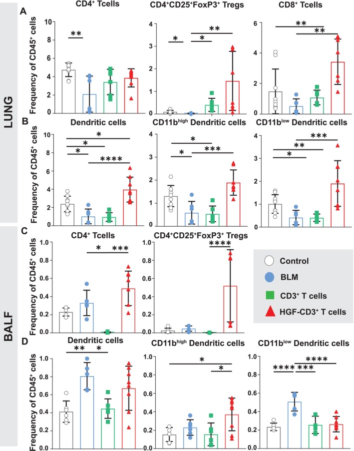
Results: Our findings demonstrate the successful T cell therapy of bleomycin-induced lung injury through the adoptive transfer of HGF-transfected T cells in mice. This treatment resulted in decreased collagen deposition and a balancing of immune cell exhaustion and cytokine homeostasis compared with untreated controls. In vitro testing showed enhanced apoptosis in myofibroblasts induced by HGF-overexpressing T cells.

Conclusions: Taken together, our data highlight the great potential of adoptive T cell transfer as an emerging therapy to counteract lung fibrosis.

Keywords: T cells homeostasis; adoptive transfer; bleomycin lung injury and fibrosis; immune exhaustion.


Conflict of interest statement

The authors declare that the research was conducted in the absence of any commercial or financial relationships that could be construed as a potential conflict of interest.

Figures

FIGURE 1
FIGURE 1
Outline of BLM mouse model, histology, fibrosis scoring, and pulmonary collagen content in the course of fibrosis. (A) Mice were either instilled intratracheal with 50 μL of saline (control), or with BLM (1.52 U/kg) and sacrificed on Day 7, Day 10, and Day 14 following instillation. (B) H&E‐stained lung sections of mice treated as outlined above. (C) Ashcroft's scoring done on H&E‐stained lung sections. (D) Lung collagen content measured by hydroxyproline assay on lung homogenate. Mice treated as outlined under A. Data show mean ± SEM, n = 12 mice per group (one point represents two pooled mice).
FIGURE 2
FIGURE 2
Frequencies of T cell and DC subsets in lung and BALF following BLM instillation. (A) Frequency of lung CD4+, Treg (CD4+CD25+FoxP3+), and CD8+ T cells within total CD45+ cells. Mice were either instilled intratracheal with BLM (1.52 U/kg) or with 50 μL of saline (control) and sacrificed on Day 7, Day 10, and Day 14 following instillation. (B) Frequency of total lung DC and CD11bhigh and CD11blow subsets. (C) BALF CD4+ and Treg (CD4+CD25+FoxP3+). (D) BALF total DC (CD11C+MHCIIhigh) and CD11bhigh and CD11blow subsets relative to total CD45+ cells. All Groups received treatment as outlined under A. Data show mean ± SEM. Statistical significance was determined by a one‐way ANOVA followed by Tukey's multiple comparison post‐test. Values were considered significantly different comparing treatments with the control group, and significances reported as p < 0.05 (*), p < 0.05 (**), p < 0.005 (***), and p < 0.0005 (****), n = 12 mice per group (one point represents two pooled mice).
FIGURE 3
FIGURE 3
Treatment approach employing nontransfected CD3 + T cells with resulting histology and pulmonary collagen content. (A) Timeline of treatment employing HGF‐CD3+ T cells. Mice were either administered intratracheal with BLM (1.52 U/kg) or with 50 μL of saline (control), and on Day 7, mice were treated either with HGF‐CD3+ T cells or nontransfected CD3+ T cells and sacrificed 7 days later (BLM group = Day 14). (B) Histologic changes in lung tissue following treatment with HGF‐transfected or nontransfected CD3+ T cells in comparison to the BLM group and control were analyzed with H&E staining (C) Ashcroft's scoring done on H&E‐stained mouse lung sections (D) Level of hepatocyte growth factor (HGF) measured in lung tissue by ELISA. (E) Total collagen content in the lung measured by hydroxyproline assay. Control = grey, BLM (Day 14) = light blue, nontransfected CD3+ T cell treatment = green. Data show mean and SEM, n = 8–12 mice per group, ****p < 0.00005, ***p < 0.0005, **p < 0.005, *p < 0.05.
FIGURE 4
FIGURE 4
Immunofluorescence staining in precision‐cut lung slices (PCLS) from BLM‐treated mice instilled with HGF‐CD3 + T cells imaged with confocal laser scanning microscopy. Mice were treated with BLM. Seven days after BLM treatment, DiO‐labeled HGF‐CD3+ T cells were instilled intratracheal, and mice were sacrificed 24 h later. Each image shows confocal optical sections of the xy‐projection (large upper image), the xz‐projection (small lower image), and the yz‐projection (small right image). Blue: actin+ filaments, red: CD3+ T cells (arrowheads), green: DiO‐stained HGF‐CD3+ T cells (squares), yellow: co‐localization DiO dye and CD3+ cells (asterisks), A = alveolus. PCLS were scanned with a confocal microscope (Zeiss, LSM 980).
FIGURE 5
FIGURE 5
Frequencies of T cell and DC subsets in the lung and BALF of BLM treated mice following treatment with HGF‐CD3 + T and nontransfected CD3 + T cells. (A) Frequency of CD4+, Treg (CD4+CD25+FoxP3+), and CD8+ T cells within total CD45+ of lung parenchyma. On Day 0, mice were either instilled intratracheal with BLM (1.52 U/kg) or with 50 μL of saline (control). Seven days after BLM administration, mice were treated with either HGF‐CD3+ T cells or nontransfected CD3+ T cells or left nontreated and sacrificed 7 days later (BLM Day 14). (B‐D) Frequency of total lung DC (CD11C+MHCIIhigh), CD11bhigh and CD11blow subsets (B), total BALF CD4+ T cells, and Treg (CD4+CD25+FoxP3+) (C) and total BALF DC (CD11C+MHCIIhigh), CD11bhigh and CD11blow subsets (D). Data show mean and SEM, n = 8–12 mice per group, ****p < 0.00005, ***p < 0.0005, **p < 0.005, *p < 0.05.
FIGURE 6
FIGURE 6
Relative mRNA expression of key inflammatory and regulatory markers, following treatment with T cells in the course of BLM‐induced lung injury. Relative mRNA expression levels measured by real‐time qPCR in lung tissue homogenates relative to nontreated control group. Relative mRNA expression of (A) HGF, (B) TGF‐β, (C) PTGES, (D) IL‐10, (E) IL‐13, and (F) IL‐17 in lung tissue normalized to the nontreated control group (i.t. instillation of 50‐μL saline) using housekeeping gene RPLP0. Data show mean and SEM, n = 4–6 mice per group, ****p < 0.00005, ***p < 0.0005, **p < 0.005, *p < 0.05.
FIGURE 7
FIGURE 7
Effect of HGF‐transfected and nontransfected T cells on TGF‐β–treated fibroblasts in vitro. (A) Frequency of total Collagen1α+Desmin+ cells; (B) frequency of total Collagen1α+Desmin+αSMA+ cells; (C) frequency of total Collagen1α+Desmin+αSMA+Caspase3+ cells; (D) frequency of total Collagen1α+Desmin+CD140α+ cells; (E) frequency of total Collagen1α+Desmin+CD140α+αSMA cells; (F) frequency of total Collagen1α+Desmin+CD140α+Caspase3+ cells. Twenty‐five thousand primary murine fibroblasts were seeded per well and treated with 8 ng/μL of TGF‐β for 24 h. Fifty thousand of nontransfected, HGF‐transfected or nontransfected CD3+ T cells were added for another 24 h before flow cytometry analysis. Control = TGF‐β–treated fibroblasts. Data show mean + SEM, n = 4–5, *p < 0.05, **p < 0.005, ***p < 0.0005, ****p < 0.00005.

References

    1. Gross T. J. and Hunninghake G. W., “Idiopathic Pulmonary Fibrosis,” New England Journal of Medicine 345 (2001): 517–525, 10.1056/NEJMRA003200. - DOI - PubMed
    1. Lee A. S., Mira‐Avendano I., Ryu J. H., and Daniels C. E., “The Burden of Idiopathic Pulmonary Fibrosis: An Unmet Public Health Need,” Respiratory Medicine 108 (2014): 955–967, 10.1016/J.RMED.2014.03.015. - DOI - PubMed
    1. Navaratnam V., Fleming K. M., West J., et al., “The Rising Incidence of Idiopathic Pulmonary Fibrosis in the UK,” Thorax 66 (2011): 462–467, 10.1136/THX.2010.148031. - DOI - PubMed
    1. Finnerty J. P., Ponnuswamy A., Dutta P., Abdelaziz A., and Kamil H., “Efficacy of Antifibrotic Drugs, Nintedanib and Pirfenidone, in Treatment of Progressive Pulmonary Fibrosis in Both Idiopathic Pulmonary Fibrosis (IPF) and Non‐IPF: A Systematic Review and Meta‐Analysis,” BMC Pulmonary Medicine 21 (2021): 411, 10.1186/S12890-021-01783-1. - DOI - PMC - PubMed
    1. Checa M., Hagood J. S., Velazquez‐Cruz R., et al., “Cigarette Smoke Enhances the Expression of Profibrotic Molecules in Alveolar Epithelial Cells,” PLoS ONE 11 (2016): e0150383, 10.1371/JOURNAL.PONE.0150383. - DOI - PMC - PubMed

MeSH terms