. 2025 Apr;29(7):e70511.
doi: 10.1111/jcmm.70511.

Unveiling the Angiogenic Potential and Functional Decline of Valve Interstitial Cells During Calcific Aortic Valve Stenosis Progression

Affiliations

Unveiling the Angiogenic Potential and Functional Decline of Valve Interstitial Cells During Calcific Aortic Valve Stenosis Progression

Adeline Blandinières et al. J Cell Mol Med. 2025 Apr.

Abstract

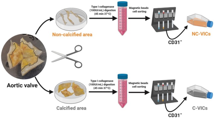
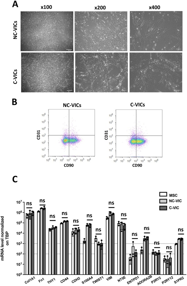
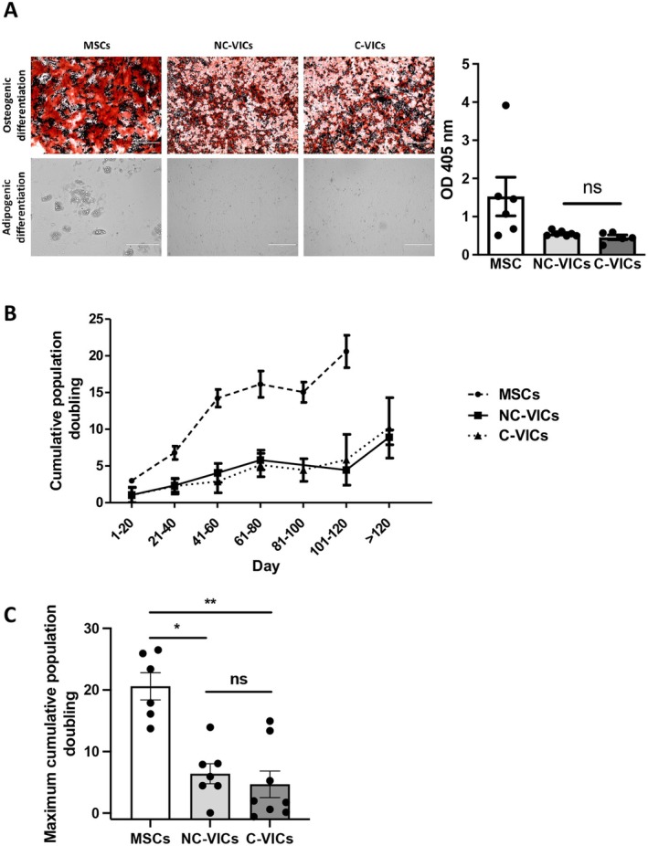
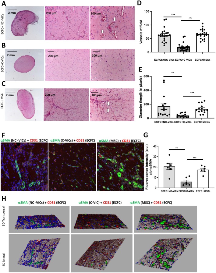
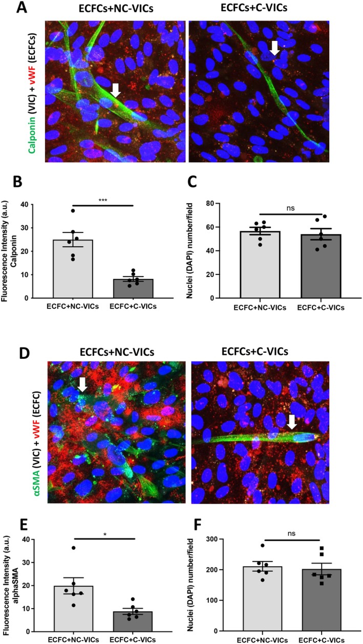
Valve interstitial cells (VICs) play a critical role in aortic valve calcification and angiogenic processes associated with calcific aortic valve stenosis (CAVS). Within the same valve, VICs from differently calcified regions can exhibit diverse phenotypic and functional properties. We hypothesised that VICs isolated from noncalcified (NC-VICs) and calcified (C-VICs) areas of human aortic valves possess distinct angiogenic characteristics. In this study, we isolated C-VICs and NC-VICs from 23 valves obtained after aortic valve replacement due to CAVS. Both VIC types exhibited similar phenotypes in culture, characterised by morphology, expression of mesenchymal/fibroblastic markers, proliferation and osteogenic differentiation. No significant differences were observed in the secretion of angiogenic factors, including VEGF-A, Ang-1, Ang-2, PlGF, bFGF between NC-VICs and C-VICs. However, when co-injected with endothelial colony-forming cells (ECFCs) into Matrigel implants in vivo in mice, implants containing NC-VICs showed significantly higher microvessel density compared to those with C-VICs (p < 0.001). Additionally, NC-VICs co-cultured with ECFCs expressed significantly higher levels of the perivascular markers αSMA and calponin compared to C-VICs (p < 0.001 and p < 0.05, respectively). In conclusion, our study reveals the heterogeneity in VIC plasticity within the aortic valve during CAVS. The diminished capacity of VICs from calcified areas to differentiate into perivascular cells suggests a loss of function as valve disease progresses. Furthermore, the ability of VICs to undergo perivascular differentiation may provide insights into valve homeostasis, angiogenesis and the exacerbation of calcification.

Keywords: aortic valve stenosis; endothelial progenitor cells; neovascularisation; perivascular cells; valve interstitial cell.


Conflict of interest statement

The authors declare no conflicts of interest.

Figures

FIGURE 1
FIGURE 1
Isolation process of VICs from calcified (C‐VICs) and noncalcified (NC‐VICs) area of the aortic valve. Noncalcified and calcified areas shown are representative of the complete cohort.
FIGURE 2
FIGURE 2
Characterisation of NC‐VICs and C‐VICs. (A) Morphology. Phase‐contrast image of VICs isolated from NC‐VICs and C‐VICs in culture at different magnifications (×100, ×200, ×400). (B) CD90 and CD31 expression in acoustic cytometry by NC‐VICs and C‐VICs (n = 6) (C) Col1A1, Fn1, THY 1, CD44, CDH2, S100A4, TWIST1, VIM, NT5E, ENTPD1, ADORA2B, P2RY2, P2RY12, S1PR2 mRNA expression by MSCs, C‐VICs and NC‐VICs (n = 3, mean ± SEM). ns, p > 0.05.
FIGURE 3
FIGURE 3
Functional characterisation of NC‐VICs and VICs. (A) Osteogenic differentiation of MSC, VICs isolated from NC‐VICs and C‐VICs revealed by Alizarin Red staining. Adipogenic differentiation of MSCs revealed by the presence of lipid drops on phase‐contrast image (scale 400 μm). Quantification of osteogenic differentiation of MSCs, NC‐VICs and C‐VICs (OD 405 nm, n = 5 to 7) (B) Cumulative population doubling of MSC, NC‐VICs and C‐VICs (n = 4). C. Maximum cumulative population doubling of MSC, NC‐VICs and C‐VICs (n = 4). ns, p > 0.05, *p < 0.05 and, **p < 0.01.
FIGURE 4
FIGURE 4
Blood vessels infiltration evaluated by haematoxylin and eosin (H&E) staining. (A) Matrigel plug of ECFCs+NC‐VICs at lower (1,25× left pannel) and higher magnification (10× central, 20× right panel) (B) Matrigel plug of ECFCs+C‐VICs at lower and higher magnification (same order) (C) Matrigel plug of ECFCs+MSCs at lower and higher magnification (same order). Arrows indicate functional vessels containing erythrocytes. (D) Vessels number quantification (n = 10 matrigel plugs for each group) (E) Diameter quantification of n = 10 Matrigel plugs for each group (A–C). **p < 0.01 and ***p < 0.001. (F) Matrigel plug section of ECFCs+NC‐VICs, ECFCs+C‐VICs, ECFCs+MSCs (from left to right). CD31 stain (red) was performed to identify endothelial cells while perivascular cells are stained by αSMA (green). (G) Quantification of αSMA stain in n = 10 different Matrigel plugs for the 3 different groups mentioned (H) 3D reconstruction of the confocal images proposed in (F).
FIGURE 5
FIGURE 5
Co‐culture of ECFCs + NC‐VICs, ECFCs + C‐VICs (A) Co‐culture of ECFCs+NC‐VICs and ECFCs+C‐VICs (400× magnification, zoom), ECFCs are positive for von Willebrand factor (vWF) stain (red) and NC‐VICs or C‐VICs are stained for calponin (green) (B) Quantification of calponin stain in n = 3 different co‐culture in duplicate (C) Quantification of nuclei in co‐culture of ECFCs+NC‐VICs and ECFCs+C‐VICs stained by calponin (D) Co‐culture of ECFCs+NC‐VICs and ECFCs+C‐VICs (400× magnification, zoom), ECFCs are positive for vWF stain (red) and NC‐VICs or C‐VICs are stained for αSMA (green). (E) Quantification of αSMA stain in n = 3 different co‐culture in duplicate (F) Quantification of nuclei in co‐culture of ECFCs+NC‐VICs and ECFCs+C‐VICs stained by αSMA. *p < 0.05 and ***p < 0.001.

Similar articles

References

    1. Lindman B. R., Clavel M.‐A., Mathieu P., et al., “Calcific Aortic Stenosis,” Nature Reviews. Disease Primers 2 (2016): 16006. - PMC - PubMed
    1. Büttner P., Feistner L., Lurz P., Thiele H., Hutcheson J. D., and Schlotter F., “Dissecting Calcific Aortic Valve Disease‐The Role, Etiology, and Drivers of Valvular Fibrosis,” Frontiers in Cardiovascular Medicine 8 (2021): 660797. - PMC - PubMed
    1. Yadgir S., Johnson C. O., Aboyans V., et al., “Global, Regional, and National Burden of Calcific Aortic Valve and Degenerative Mitral Valve Diseases, 1990–2017,” Circulation 141 (2020): 1670–1680. - PubMed
    1. Rajamannan N. M., Nealis T. B., Subramaniam M., et al., “Calcified Rheumatic Valve Neoangiogenesis Is Associated With Vascular Endothelial Growth Factor Expression and Osteoblast‐Like Bone Formation,” Circulation 111 (2005): 3296–3301. - PMC - PubMed
    1. Syväranta S., Helske S., Laine M., et al., “Vascular Endothelial Growth Factor–Secreting Mast Cells and Myofibroblasts,” Arteriosclerosis, Thrombosis, and Vascular Biology 30 (2010): 1220–1227. - PubMed

MeSH terms

Supplementary concepts

LinkOut - more resources