

Enhancing bone radiology images classification through appropriate preprocessing: a deep learning and explainable artificial intelligence approach
- PMID: 40160653
- PMCID: PMC11948367
- DOI: 10.21037/qims-24-1745
Enhancing bone radiology images classification through appropriate preprocessing: a deep learning and explainable artificial intelligence approach
Abstract
Background: Medical image classification has been an important application for deep learning techniques for over a decade, and since the emergence of explainable artificial intelligence (XAI), researchers have started using XAI to validate the results produced by these black box models. In the research field, it has become clear that accuracy and efficiency are not the only crucial factors for developing medical deep learning models; the authenticity of results and the accountability of the model and its creator also matter greatly. The objective of this study is to emphasize the importance of authenticity of the results and the accountability of deep learning models used for medical purposes, through proposing targeted preprocessing method for medical dataset processed by deep learning models.
Methods: In this paper we conduct comparison experiments on processing two bone radiology image datasets using various deep learning neural networks, while emphasizing on the effect of appropriate preprocessing methods for the dataset towards the models' prediction performance. Comparisons are conducted both horizontally, between performance of different neural networks; and vertically, of using same models processing datasets before and after going through appropriate preprocessing procedures. Furthermore, we evaluate the experimental results not only quantitatively, but also visually by using XAI techniques, in order to determine the reasonability and reliability of the predictions from the experiments.
Results: Results showed that for the bone radiology image dataset used for our experiment, among the five comparison models, DenseNet201 achieved the highest validation accuracy of 78%. Using the same models to process the abovementioned dataset after conducting appropriate preprocessing procedures, performance for all models have increased by an average of 0.06. Using XAI technique to evaluate the comparison results for before/after preprocessing experiments, we could observe that the appropriate preprocessing method effectively helped the models to concentrate on the abnormality areas on the radiology images comparing to processing raw images.
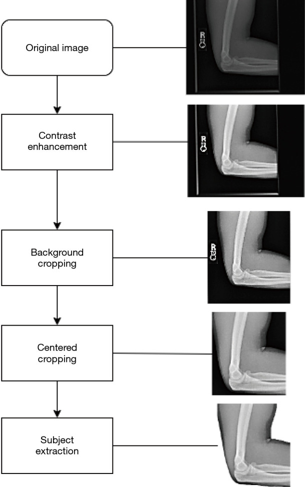
Conclusions: The novelty of this paper lies in its specific application of extended preprocessing techniques-namely, the removal of background and irrelevant parts-to medical images for improving the performance of deep learning models in classification tasks. While the concept of preprocessing images has been explored by many researchers, applying such targeted preprocessing steps to medical images, combined with the use of XAI to validate and illustrate the benefits, is a novel approach. This paper highlights the unique requirements of medical image data and proposes an innovative method to enhance model accuracy and reliability in medical diagnostics by removing background and redundant features from the images.
Keywords: Bone abnormality; convolutional neural network (CNN); deep learning; explainable artificial intelligence (XAI); medical image preprocessing.
Copyright © 2025 AME Publishing Company. All rights reserved.
Conflict of interest statement
Conflicts of Interest: All authors have completed the ICMJE uniform disclosure form (available at https://qims.amegroups.com/article/view/10.21037/qims-24-1745/coif). The authors have no conflicts of interest to declare.
Figures


References
-
- Gharaibeh M, Almahmoud M, Ali MZ, Al-Badarneh A, El-Heis M, Abualigah L, Altalhi M, Alaiad A, Gandomi AH. Early Diagnosis of Alzheimer’s Disease Using Cerebral Catheter Angiogram Neuroimaging: A Novel Model Based on Deep Learning Approaches. Big Data Cogn. Comput 2022;6:2.
LinkOut - more resources
Full Text Sources