The Development of the Umbilical Vein and Its Anatomical and Clinical Significance
- PMID: 40161047
- PMCID: PMC11954436
- DOI: 10.7759/cureus.79712
The Development of the Umbilical Vein and Its Anatomical and Clinical Significance
Abstract
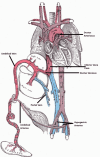
The umbilical vein is one of the most essential vessels in the human embryo. Anatomical structures though may vary in several cases. During the fourth and eighth weeks of gestation, the umbilical cord is formed. Initially, two umbilical arteries and veins exist. During development, the obliteration of the right umbilical vein occurs. The fetus and its liver receive macronutrients and oxygen from the placenta via the umbilical vein, which primarily supplies the left lobe of the liver before branching into the left portal vein and the ductus venosus. The ductus venosus directs blood from the umbilical vein directly into the systemic circulation through the inferior vena cava and right atrium, bypassing the fetal liver. In some cases, variations are observed. Disorders of the umbilical veins may involve the persistence of embryological structures, abnormal insertion or course, and the presence of supernumerary vessels. For example, the persistence of the right umbilical vein, duplication of the umbilical vein, and umbilical vein varix are some important variations to acknowledge in order to be able to understand the potential outcomes of the newborn. The majority of venous system anomalies are rare, and some may remain completely asymptomatic. Different forms of umbilical cord abnormalities, however, may be potentially fatal or pose a serious threat to fetal health. Therefore, clinically, early detection of these malformations is highly important in order to make a proper diagnosis and management of care. The aim of this study is to acknowledge the different types of umbilical vein variations through its development and its relation with liver parenchyma in order to achieve a better understanding and planning in surgical interventions. An advanced review search of the literature was undertaken. The literature review was conducted using the search engine of the PubMed database and Google Scholar. The years included in data collection were 1960-2022. All articles that met the inclusion criteria were taken under consideration.
Keywords: anatomical variations of liver vasculature; development of umbilical vein; liver surgery; umbilical vein and surgery; umbilical vein variations.
Copyright © 2025, Kozadinos et al.
Conflict of interest statement
Conflicts of interest: In compliance with the ICMJE uniform disclosure form, all authors declare the following: Payment/services info: All authors have declared that no financial support was received from any organization for the submitted work. Financial relationships: All authors have declared that they have no financial relationships at present or within the previous three years with any organizations that might have an interest in the submitted work. Other relationships: All authors have declared that there are no other relationships or activities that could appear to have influenced the submitted work.
Figures


References
-
- Cochard LR, Netter FH. Teterboro, NJ : Icon Learning Systems. Teterboro (NJ): Icon Learning Systems; 2002. Netter's atlas of human embryology.
-
- Edinburgh: Churchill Livingstone/Elsevier; 2008. Gray's anatomy: the anatomical basis of clinical practice.
-
- Blackburn ST. Philadelphia (PA): Saunders; 2017. Maternal, fetal, & neonatal physiology: a clinical perspective.
-
- Norton ME, Scoutt LM, Feldstein VA, Callen PW. Philadelphia (PA): Elsevier; 2017. Callen's ultrasonography in obstetrics and gynecology.
-
- CT in lobar atrophy of the liver caused by alveolar echinococcosis. Rozanes I, Acunaş B, Celik L, Minareci O, Gökmen E. J Comput Assist Tomogr. 1992;16:216–218. - PubMed
Publication types
LinkOut - more resources
Full Text Sources
Miscellaneous
