

Irreducible Isolated Radial Head Dislocation in a Child Due to Annular Ligament Interposition: A Case Report
- PMID: 40161065
- PMCID: PMC11952084
- DOI: 10.7759/cureus.79694
Irreducible Isolated Radial Head Dislocation in a Child Due to Annular Ligament Interposition: A Case Report
Abstract
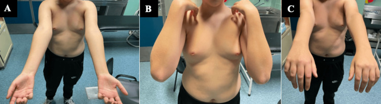
The annular ligament of the elbow is essential for its stability, playing a key role in both the proximal radioulnar and humeroradial joints, as well as supporting surrounding muscles and ligaments. Radial head dislocation is rare in children, and when isolated they can be challenging to reduce and may require surgical intervention. An 11-year-old boy presented with an anteromedial dislocation of the radial head after a fall. Initial, closed reduction was attempted and failed, requiring surgical intervention, where we found a rupture of the annular ligament and interposition, which was repaired after reduction. At one year of follow-up, the patient achieved full range of motion and stability of the elbow. Radial head dislocation is rare in children and even more rare without associated ulna fractures. Evaluating radiographs for plastic deformities of the ulna is crucial, as these injuries are often overlooked. Failed closed reductions may rise suspicion of interposed structures. Isolated post-traumatic radial head dislocation is a rare occurrence requiring prompt recognition and management. While most cases are treated with closed reduction, those that are not reducible or suspected soft tissue interposition may require open reduction. Thorough clinical evaluation, both vascular and neurologic and preoperative imaging are essential. Early intervention and meticulous surgical techniques can lead to favorable functional outcomes.
Keywords: annular ligament; irreducible radial head; pediatric elbow trauma; radial head dislocation; radial head open reduction.
Copyright © 2025, Fabião et al.
Conflict of interest statement
Human subjects: Consent for treatment and open access publication was obtained or waived by all participants in this study. Conflicts of interest: In compliance with the ICMJE uniform disclosure form, all authors declare the following: Payment/services info: All authors have declared that no financial support was received from any organization for the submitted work. Financial relationships: All authors have declared that they have no financial relationships at present or within the previous three years with any organizations that might have an interest in the submitted work. Other relationships: All authors have declared that there are no other relationships or activities that could appear to have influenced the submitted work.
Figures


References
-
- The annular ligament: an anatomical study. Bozkurt M, Acar HI, Apaydin N, Leblebicioglu G, Elhan A, Tekdemir I, Tonuk E. Am J Sports Med. 2005;33:114–118. - PubMed
-
- Nursemaid's elbow: radial head subluxation injuries in children. Paluch LK. JAAPA. 2024;37:18–21. - PubMed
-
- Treatment of chronic isolated radial head dislocation in children. De Boeck H. Clin Orthop Relat Res. 2000:215–219. - PubMed
Publication types
LinkOut - more resources
Full Text Sources