[Preprint]. 2025 Mar 14:2025.03.11.642495.
doi: 10.1101/2025.03.11.642495.

Characterization of MEK1/2 Degraders Uncovers a Kinase-Independent Role for MEK1/2 in the Stabilization and Maturation of CRAF

Affiliations

Characterization of MEK1/2 Degraders Uncovers a Kinase-Independent Role for MEK1/2 in the Stabilization and Maturation of CRAF

Jason S Wasserman et al. bioRxiv. .

Abstract

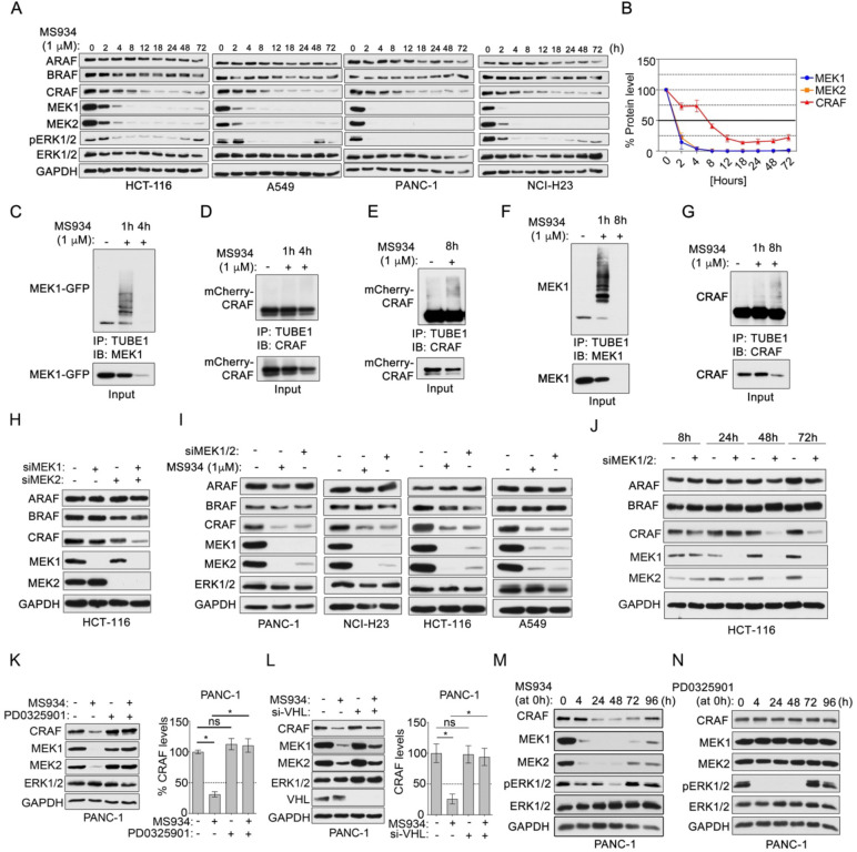
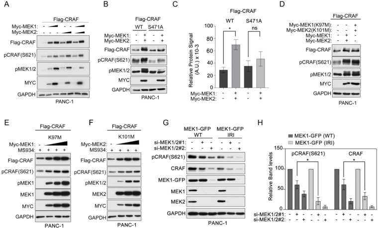
Altered MAPK signaling frequently occurs in human disease. MEK1 and MEK2 (MEK1/2) are central protein kinases in the MAPK signaling cascade that phosphorylate ERK1/2 promoting cell growth. MEK1/2 degraders offer a strategy to characterize both kinase-dependent and independent functions of MEK1/2. Here, we discovered that MEK1/2 degradation, but not kinase inhibition, caused the subsequent degradation of upstream kinase CRAF via a cell-intrinsic mechanism. MEK1/2 binding to CRAF, but not MEK1/2 catalytic activity, was required for CRAF protein stability and maturation to a functional kinase. In the absence of MEK1/2, a minor pool of newly synthesized immature CRAF that had anti-apoptotic functions remained. Finally, we showed that a stable primed CRAF-MEK1/2 signaling complex existed in cells that required RAS binding to potentiate MEK-ERK phosphorylation. Together, we've discovered a previously unrecognized kinase-independent function of MEK1/2, while contextualizing MEK1/2 as an integral component of the CRAF activation cycle beyond the conventional CRAF-MEK kinase-substrate paradigm.


Conflict of interest statement

Competing interests: J.S.D. is an inventor on patent application 63/447,909 “Methods of Degrading Raf (RAF) Protein In Cells Using Mitogen-Activated Protein Kinase Kinase 1/2 (MEK1/2) Protein Degraders”. J.J. and J. H. are inventors of a patent application filed by Icahn School of Medicine at Mount Sinai. The Jin laboratory received research funds from Celgene Corporation, Levo Therapeutics, Cullgen Inc. and Cullinan Oncology. J.J. is a cofounder and equity shareholder in Cullgen Inc. and a consultant for Cullgen Inc., EpiCypher Inc., and Accent Therapeutics Inc. The other authors declare that they have no competing interests related to this project.

Figures

Figure 1.
Figure 1.. MEK1/2 degradation but not inhibition promotes CRAF loss.
(A-B) Volcano plot of proteins increased or reduced in abundance in PANC-1 cells treated with PD0325901 (A) or MS934 (B) for 24 hours relative to DMSO-treated cells. Differences in protein log2 LFQ intensities among treated or control cells were determined by paired t-test permutation-based adjusted P values at FDR of <0.05 using Perseus software. (C-D) Immunoblotting for MAPK signaling components in PANC-1 cells treated with escalating doses of PD0325901, MS934, or MS934(−) for 24 hours. Blots are representative, and densitometric analysis of CRAF (D) are means ± SEM from three blots, each normalized to the loading control, GAPDH. (E) ARAF, BRAF, RAF1 mRNA levels in PANC-1 cells following treatment with 1 μM MS934 for 24 hours, as determined by qRT-PCR. Data are means ± SEM of three independent experiments. (F) Immunoblotting for CRAF, MEK1/2, and ERK1/2 in PANC-1 cells treated with DMSO, 0.1 μM MS934, 0.1 μM bortezomib, or the combination for 24 hours. Cells were treated with bortezomib 2 hours prior to administering MS934. Blots are representative, and densitometric analysis of CRAF protein levels are means ± SEM from three blots, each normalized to the loading control, GAPDH. (G) Immunoblotting for MAPK signaling components in non-tumorigenic cell line MCF-10A treated with escalating doses of PD0325901, or MS934 for 24 hours. (H) Immunoblotting for MAPK signaling components in A549 cells treated with 1 μM MS934, MEK1/2 inhibitors, or ERK1/2 inhibitor for 24 hours. (I-J) Microscopy image (I) and immunoblot analysis (J) of mCherry-CRAF or MEK1-GFP expression in PANC-1 cells treated with 1 μM MS934 for 24 hours. Images are representative of three independent experiments. Scale bars, 200 μm. (K) Immunoblot analysis for mCherry-CRAF and MEK1-GFP in PANC-1 cells treated with DMSO, 1 μM MS934, 0.1 μM bortezomib, or the combination for 24 hours. Cells were treated with bortezomib 2 hours prior to administering MS934. (L-M) Immunoblot analysis of MAPK signaling components in Capan-1 cells treated with escalating doses of MS432 (H) or MS910 (I) for 24 hours.
Figure 2.
Figure 2.. CRAF protein stability dependent on MEK1/2 expression in cells
(A-B) Immunoblot analysis of MAPK signaling in HCT-116, A549, PANC-1, and NCI-H23 following 1 μM MS934 treatment across a 72-hour time course. Fresh MS934 was administered every 24 hours. Blots are representative, and densitometric analysis of MEK1, MEK2, and CRAF (B) are means ± SEM across cell line blots, each normalized to the loading control, GAPDH. (C-G) Immunoprecipitation (IP) of ubiquitin-modified proteins using TUBE1 beads from PANC-1 cells co-expressing mCherry-CRAF and MEK1-GFP (C-E) or HCT-116 cells (F-G) treated with 1 μM MS934 for 1, 4 or 8 hours. The TUBE1 IP elutes were blotted with indicated antibodies. (H) Immunoblot analysis of RAF and MEK1/2 protein levels in HCT-116 cells treated with control siRNAs or MEK1, MEK2, or MEK1 and MEK2 siRNAs and cultured for 72 hours. (I) Immunoblot analysis of RAF and MEK1/2 levels in PANC-1, NCI-H23, HCT-116 and A549 treated with 1 μM MS934 or MEK1 and MEK2 siRNAs (siMEK1/2). (J) Immunoblot analysis of RAF and MEK1/2 levels in HCT-116 cells treated with siRNAs targeting both MEK1 and MEK2 for 8, 24, 48, and 72 hours. (K) Immunoblotting for CRAF, MEK1/2, or ERK1/2 in PANC-1 cells treated with DMSO, 1 μM PD0325901, 0.1 μM MS934, or the combination for 24 hours. Cells were treated with PD0325901 2 hours prior to adding MS934. Blots are representative, and densitometric analysis of CRAF levels are means ± SEM from three blots, each normalized to the loading control, GAPDH. (L) Immunoblot analysis for CRAF, MEK1/2 or ERK1/2 in PANC-1 cells transfected with control siRNA or siRNAs targeting VHL and treated with DMSO or 0.1 μM MS934 for 24 hours. Blots are representative, and densitometric analysis of CRAF protein levels are means ± SEM from three blots, each normalized to the loading control, GAPDH. (M-N). Immunoblot analysis of MAPK signaling in PANC-1 cells treated with a single dose of 0.1 μM MS934 (M) or PD0325901 (N) and followed across a 96-hour period. No drug replenishment was performed.
Figure 3.
Figure 3.. MEK1/2 degradation or knockdown caused rapid dephosphorylation of CRAF but not ARAF or BRAF, promoting protein degradation.
(A-B) Immunoblot analysis of CRAF phosphorylation in HCT-116, A549, PANC-1, and NCI-H23 following 1 μM MS934 treatment across a 72-hour time course. Fresh MS934 was administered every 24 hours. Blots are representative, and densitometric analysis of CRAF total, p-S259, p-S289/296/301, p-S338, and p-S621 (B) are means ± SEM across cell line blots, each normalized to the loading control, GAPDH. CRAF, MEK1, and MEK2 blots are also shown in Fig. 2A. (C) Immunoblotting for CRAF phosphorylation in HCT-116 cells treated with 1 μM PD0325901, MS934, or the combination for 0, 0.5, 1, 2, 3, 4, or 24 hours. (D) Immunoblot analysis of CRAF phosphorylation levels in HCT-116 cells treated with non-targeting siRNAs or MEK1, MEK2, or MEK1 and MEK2 siRNAs and cultured for 48 hours. (E) Immunoblot analysis of CRAF phosphorylation levels in HCT-116 cells treated with siRNAs targeting both MEK1 and MEK2 for 8, 24, 48, and 72 hours. (F-G). Immunoblotting of CRAF phosphorylation at S621 levels in HCT-116 cells treated with 1 μM MS934 alone or in combination with 2.5 nM Calyculin A for 0, 1, or 2 hours. Cells were pre-treated with Calyculin A for 30 minutes followed by MS934 treatment for indicated times. Blots are representative, and densitometric analysis of CRAF p-S621 (G) are means ± SD of 3 independent blots, each normalized to total CRAF protein and the loading control, GAPDH. (H-I) Immunoprecipitation (IP) of endogenous CRAF from HCT-116 cells treated with 1 μM PD0325901 (H) or 1μM MS934 for 0, 4, or 24 hours. The CRAF IPs were blotted with the indicated antibodies. Input samples were used as controls. (J-L) Immunoblot analysis of RAF phosphorylation in PANC-1 cells expressing Flag-CRAF(J), Flag-BRAF (K), or Flag-ARAF (L) treated with 0, 0.1, and 1 μM MS934 for 24 hours. (M-O) Immunoprecipitation (IP) of endogenous CRAF (M), BRAF (N), or ARAF (O) from HCT-116 cells treated with 1 μM MS934 for 0, 4, or 24 hours. The RAF IPs were blotted with the indicated antibodies. Input samples were used as controls.
Figure 4.
Figure 4.. MEK1/2-binding to CRAF and not catalytic activity promotes CRAF stabilization and activation.
(A) Immunoblot analysis of total and phosphorylated Flag-CRAF levels in PANC-1 cells transfected with Flag-CRAF (2.5 μg) and escalating concentrations of Myc-MEK1 (37.5, 75 ng), Myc-MEK2 (50, 100ng) or the combination of Myc-MEK1 and Myc-MEK2 (halving amounts) for 48 hrs. (B-C) Immunoblot analysis of total and phosphorylated Flag-CRAF levels in PANC-1 cells transfected with Flag-CRAF or Flag-CRAF(S471A) (2.5 μg) and Myc-MEK1 (37.5 ng)/ Myc-MEK2 (25 ng). Blots are representative, and densitometric analysis of p-S621 and CRAF (C) are means ± SD from three blots, each normalized to the loading control, GAPDH. (D) Immunoblot analysis of total and phosphorylated Flag-CRAF levels in PANC-1 cells transfected with Flag-CRAF and Myc-MEK1/2 wild-type of Myc-MEK1(K97M)/MycMEK2(K101M) (25 ng per). (E-F). Immunoblotting of total and phosphorylated Flag-CRAF levels in MS934-treated (1 μM) PANC-1 cells transfected with Flag-CRAF (2.5 μg) and escalating concentrations of Myc-MEK1(K97M) (75, 150, 300 ng) (E) or Myc-MEK2(K101M) (75, 150, 300 ng) (F) for 48 hrs. Cells were pre-treated with MS934 for 4 hours to deplete endogenous MEK1/2. (G-H) Immunoblot analysis of endogenous CRAF total and p-S621 levels in PANC-1 cells expressing si-MEK1/2#1 resistant MEK1-GFP(WT) o MEK1-GFP(IRI) treated with control, si-MEK1/2#1 or MEK1/2#2 siRNAs for 72 hours. Blots are representative, and densitometric analysis of p-S621 and CRAF (G) are means ± SEM from three blots, each normalized to the loading control, GAPDH.
Figure 5.
Figure 5.. MEK1/2 is required for stabilizing mature CRAF but not the immature form
(A) Immunoblot analysis of CRAF protein levels in HCT-116 cells treated with MEK1/2 siRNAs for 72 hours or 1 μM MS934 for 24 hours. (B) Immunoblotting for CRAF total and p-S621 levels in HCT-116 cells treated with DMSO, 1 μM MS934, 100 μg/ml cycloheximide, 0.05 μM STA9090, or the combination of MS934 and cycloheximide. (C) Immunoblot analysis of RAF levels in HCT-116 cells treated with DMSO, 1 μM MS934, 0.05 μM STA9090, or the combination for 24 h. (D) Immunoblot analysis for p-S621 and CRAF levels in HCT-116 cells treated with DMSO or escalating doses of MS934 in the presence or absence of 2 μM RAD001 for 24 hours. (E) Immunoblotting for Flag-CRAF levels in PANC-1 cells transfected with Flag-CRAF(WT) or Flag-CRAF(S621A) and cells expressing Flag-CRAF(WT) were treated with DMSO, 0.1 or 1 μM MS934. (F-H) Immunoblot analysis of Flag-CRAF levels in PANC-1 cells stably expressing MEK1-GFP transfected with Flag-CRAF(WT) or Flag-CRAF(S621A) (F) or Flag-CRAF(WT) or Flag-CRAF(K375M) (G) or Flag-CRAF(S471A) treated with DMSO, 1 μM MS934, 100 μg/ml cycloheximide, or the combination of MS934 and cycloheximide for 24 hours. (I) Immunoblotting for Flag-CRAF levels in PANC-1 cells stably expressing MEK1-GFP transfected with Flag-CRAF(WT), Flag-CRAF(S621A), Flag-CRAF(S471A) or Flag-CRAF(K375M) treated with DMSO, or 0.05 μM STA9090 for 24 hours.
Figure 6.
Figure 6.. Immature CRAF has anti-apoptotic functions outside the MAPK signaling pathway.
(A-B) Immunoblot analysis of cleaved PARP levels in HCT-116 (A) or A549 (B) cells treated with DMSO or 1 μM MS934 or upon short hairpin RNA (shRNA)-mediated depletion of CRAF (shCRAF) or uninduced (1 μg/ml doxycycline for 120 h (A), 72 h (B). (C-D) Immunoblot analysis of cleaved PARP levels in HCT-116 (C) or A549 (D) cells treated with DMSO or 1 μM MS934 for 24 h followed by short hairpin RNA (shRNA)-mediated depletion of CRAF (shCRAF) or uninduced (1 μg/ml doxycycline for 96 h (C) or 120 h (D)). (E) Immunoprecipitation (IP) of Flag-CRAF from PANC-1 cells stably expressing MEK1-GFP treated with DMSO or 1 μM MS934 for 24 h or IP of Flag-CRAF(K375M). The Flag-CRAF IPs were blotted with the indicated antibodies. IP of Flag-GFP and input samples were used as controls. (F-G) Caspase 3/7 activation analysis in HCT-116 (F) or A549 (G) cells treated with DMSO, 0.005 μM PD0325901, 0.5 μM MS934, 1 μM Navitoclax, or the combination of PD0325901 or MS934 and Navitoclax for 24 h. Data are means ± SEM of three independent experiments. (H-I) Soft-agar colony formation assay in HCT-116 (H) or A549 (I) cells treated with DMSO, 0.005 μM PD0325901, 0.5 μM MS934, 1 μM Navitoclax, or the combination of PD0325901 or MS934 and Navitoclax for 2 weeks. Images are magnifications of representative fields. The corresponding colony counts are means ± SEM derived from multiple fields (N=6).
Figure 7.
Figure 7.. A stable complex of CRAF-MEK1/2-HSP90–14-3–3 exists that is primed for activation by RAS.
(A) Immunoprecipitation (IP) of Flag-tagged CRAF constructs from PANC-1 cells stably expressing MEK1-GFP. The Flag-CRAF IPs were blotted with the indicated antibodies. IP of Flag-GFP and input samples were used as controls. (B) Immunoblot analysis of Flag-tagged CRAF constructs in PANC-1 cells stably expressing MEK1-GFP transfected with Flag-CRAF(WT), Flag-CRAF(R401H) or Flag-CRAF(R89L) treated with DMSO, 1 μM MS934, 100 μg/ml cycloheximide, or the combination of MS934 and cycloheximide for 24 hours. (C-E) Immunoblot analysis of CRAF signaling in PANC-1 cells co-transfected with Myc-MEK1/Myc-MEK2 and Flag-CRAF(WT), Flag-CRAF(R89L), Flag-CRAF(R401H) or Flag-CRAF(K375M) for 48 hrs. Blots are representative, and densitometric analysis of p-MEK1/2 and (D/E) are means ± SEM from three blots, each normalized to the loading control, GAPDH, Myc-tag and Flag levels. (F) Immunoprecipitation (IP) of endogenous MEK1 from HCT-116 cells treated with 1 μM ERK1/2 inhibitor SCH772984 for 24 hours. The MEK1 IP was blotted for CRAF and MEK1 proteins. Blots are representative. (G) Model of MEK1/2 regulation of CRAF stabilization, maturation, and activation. Figure created in BioRender. Duncan, J. (2025) https://BioRender.com/t50o519.

References

    1. ADVANI S. J., CAMARGO M. F., SEGUIN L., MIELGO A., ANAND S., HICKS A. M., AGUILERA J., FRANOVIC A., WEIS S. M. & CHERESH D. A. 2015. Kinase-independent role for CRAF-driving tumour radioresistance via CHK2. Nat Commun, 6, 8154. - PMC - PubMed
    1. ALABI S., JAIME-FIGUEROA S., YAO Z., GAO Y., HINES J., SAMARASINGHE K. T. G., VOGT L., ROSEN N. & CREWS C. M. 2021. Mutant-selective degradation by BRAF-targeting PROTACs. Nature Communications, 12, 920. - PMC - PubMed
    1. BAHAR M. E., KIM H. J. & KIM D. R. 2023. Targeting the RAS/RAF/MAPK pathway for cancer therapy: from mechanism to clinical studies. Signal Transduct Target Ther, 8, 455. - PMC - PubMed
    1. BONED DEL RIO I., YOUNG L. C., SARI S., JONES G. G., RINGHAM-TERRY B., HARTIG N., REJNOWICZ E., LEI W., BHAMRA A., SURINOVA S. & RODRIGUEZ-VICIANA P. 2019. SHOC2 complex-driven RAF dimerization selectively contributes to ERK pathway dynamics. Proc Natl Acad Sci U S A, 116, 13330–13339. - PMC - PubMed
    1. BROMBERG-WHITE J. L., ANDERSEN N. J. & DUESBERY N. S. 2012. MEK genomics in development and disease. Brief Funct Genomics, 11, 300–10. - PMC - PubMed

Publication types