[Preprint]. 2025 Mar 12:2025.03.07.641755.
doi: 10.1101/2025.03.07.641755.

Antibody-Free Immunopeptide Nano-Conjugates for Brain-Targeted Drug Delivery in Glioblastoma Multiforme

Affiliations

Antibody-Free Immunopeptide Nano-Conjugates for Brain-Targeted Drug Delivery in Glioblastoma Multiforme

Saurabh Sharma et al. bioRxiv. .

Update in

Abstract

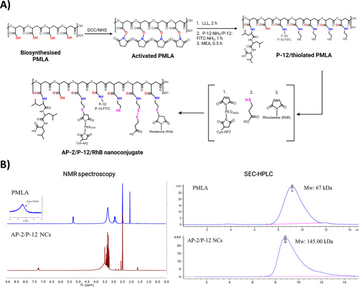
Glioblastoma Multiforme (GBM) represents a significant clinical challenge amongst central nervous system (CNS) tumors, with a dismal mean survival rate of less than 8 months, a statistic that has remained largely unchanged for decades (National Brain Society, 2022). The specialized intricate anatomical features of the brain, notably the blood-brain barrier (BBB), pose significant challenges to effective therapeutic interventions, limiting the potential reach of modern advancements in immunotherapy to impact these types of tumors. This study introduces an innovative, actively targeted immunotherapeutic nanoconjugate (P12/AP-2/NCs) designed to serve as an immunotherapeutic agent capable of traversing the BBB via LRP-1 receptor-mediated transcytosis. P12/AP-2/NCs exert its immune-modulating effects by inhibiting the PD-1/PD-L1 axis through a small-size PD-L1/PD-L2 antagonist peptide Aurigene NP-12 (P12). P12/AP-2/NCs are synthesized from completely biodegradable, functionalized high molecular weight β-poly(L-malic acid) (PMLA) polymer, conjugated with P12 and Angiopep-2 (AP2) to yield P12/AP-2/NCs. Evaluating nanoconjugates for BBB permeability and 3-D tumor model efficacy using an in vitro BBB-Transwell spheroid based model demonstrating successful crossing of the BBB and internalization in brain 3D tumor environments. In addition, the nanoconjugate mediated T cell's cytotoxicity on 3D tumor region death in a U87 GBM 3-D spheroid model. AP2/P12/NCs is selectively inhibited in PD1/PDL1 interaction on T cells and tumor site, increasing inflammatory cytokine secretion and T cell proliferation. In an in-vivo murine brain environment, rhodamine fluorophore-labeled AP2/P12/NCs displayed significantly increased accumulation in the brain during 2-6 h time intervals post-injection with a prolonged bioavailability over unconjugated peptides. AP2/P12/NCs demonstrated a safety profile at both low and high doses based on major organ histopathology evaluations. Our findings introduce a novel, programmable nanoconjugate platform capable of penetrating the BBB for directed delivery of small peptides and significant immune environment modulation without utilizing antibodies, offering promise for treating challenging brain diseases like glioblastoma multiforme and beyond.

Keywords: 3D Tumor-BBB Model; Biodistribution; Immunotherapy; Nanoconjugate.


Figures

Figure 1.
Figure 1.
The introductory mechanism of action for AP-2/P-12/RhB nanoconjugates (NCs) involves a multi-step process that enhances their therapeutic efficacy against brain tumors. In a) the AP-2 in the nanoconjugates utilize LRP1 receptor-mediated transcytosis to cross the BBB and BBTB. This process allows the NCs to reach the brain tumor site. Once at the tumor site, b) the P-12 blocks the PD-1/PD-L1 immune checkpoint interaction. This blockade occurs between T cells and PDL1-positive brain tumor cells, preventing the tumor cells from evading immune surveillance. This disruption of the PD-1/PD-L1 axis leads to enhanced T cell activity, characterized by: i) Increased T cell (CD4+/CD8+ Cells) proliferation and ii) Upregulation of major proinflammatory cytokine expression such as TNFα, IL-2, IFN-γ. The activated T cells exhibit heightened cytotoxic activity against the brain tumor cells, promoting tumor cell death and potentially reducing tumor burden.
Figure 2.
Figure 2.
Synthesis of AP-2/P-12/RhB NCs: A) Synthesis of AP2–2/P-12/Rhb NCs from PMLA which is a multistep process involving the activation of PMLA obtained from a biosynthetic source, which will render the PMLA backbone to conjugate the AP-2, P-12, and RhB moieties successfully. B) Characterization by using 1H NMR to confirm successful conjugation of functional moieties to the PMLA backbone and SEC-HPLC for determining the total molecular weight distribution of the synthesized PMLA and AP-2/P-12 NCs.
Figure 3.
Figure 3.
A) Schematic representation of a BBB-transwell model demonstrating nanoparticle receptor-mediated transcytosis and PDL1 receptor-mediated interaction with GBM spheroids wherein we have incorporated human brain microvascular endothelial cells (HBMECs) on a fibronectin layer to mimic the blood-brain barrier (BBB). The model illustrates the successful crossing of AP-2/P-12/RhB NCs through the BBB and their subsequent interaction with glioblastoma multiforme (GBM) tumor spheroids in a separate compartment. B) Cellular uptake of nanoconjugates in a BBB transwell model: Confocal laser scanning microscopy (CLSM) analysis of AP-2/P-12/RhB NCs uptake by HBMECs (seeded as a monolayer on fibronectin-coated transwells) mimicking the BBB and incubated with 0.1 mg/mL of AP-2/P-12/RhB NCs for 2 h. C) Nanoconjugate penetration in 3D GBM spheroid model carried out by CLSM visualization of AP-2/P-12/RhB NCs (100 ug/mL) uptake and distribution within 3D U87 glioblastoma spheroids after 4 h incubation at 37°C. D) Quantitative analysis of nanoconjugate uptake carried out by measuring the mean fluorescence intensity from CLSM images, representing the relative uptake and accumulation of AP-2/P-12/RhB NCs in both the BBB model and GBM spheroids. Data are presented as mean ± standard error of the mean (SEM). Statistical significance was determined using one-way ANOVA followed by post-hoc tests, with ** p < 0.01, *** p < 0.001, and **** p < 0.0001 indicating significance levels between treatment groups.
Figure 4:
Figure 4:
AP-2/P-12 nanoconjugates (NCs) modulate T lymphocyte proliferation and proinflammatory cytokine signaling. A) Schematic representation of the quantitative comparative T lymphocyte proliferation rescue analysis with Flow cytometry and ELISA. B) Subset-specific analysis of CD4+ and CD8+ T lymphocyte proliferation in response to anti-PD-1 antibody, P-12 peptide, and AP-2/P-12 NCs. Data are presented as mean ± standard error of the mean (SEM). Statistical significance was determined using one-way ANOVA followed by post-hoc tests, with ** p < 0.01, *** p < 0.001, and **** p < 0.0001 indicating significance levels between treatment groups. The results suggest a synergistic effect of the AP-2/P-12 NCs in promoting T cell activation and proliferation, which directly enhance anti-tumor immune responses.
Figure 5.
Figure 5.
AP-2/P-12 nanoconjugates (NCs) enhance T cell-mediated cytotoxicity against glioblastoma multiforme (GBM) spheroids and inhibit their growth. A) Visualization of T cell-mediated cytotoxicity: Confocal microscopy images demonstrating the cytotoxic effects of T cells on GBM spheroids in the presence of P12 peptide and AP-2/P-12 NCs. Live cells are visualized using calcein AM (green fluorescence), while dead cells are identified by ethidium bromide (EtBr) staining (red fluorescence). This dual-fluorescence assay allows for the simultaneous quantification of live and dead cells within the spheroids. This provides a direct measure of T cell-mediated cytotoxicity. B) Temporal analysis of 3D tumor spheroid growth: Quantitative assessment of GBM spheroid size over 7 days when co-cultured with T cells in the presence of P12 peptide alone or AP-2/P12 NCs using brightfield microscopy. A reduced growth rate of spheroid size in the AP-2/P-12 NC treatment group over the 4 days can be observed. Data are presented as mean ± standard error of the mean (SEM). Statistical significance was determined using one-way ANOVA followed by post-hoc tests, with ** p < 0.01, *** p < 0.001, and **** p < 0.0001 indicating significance levels between treatment groups.
Figure 6.
Figure 6.
In vivo whole body and ex vivo tissue biodistribution at 2 h and 6 h after intravenous (i.v.) administration of AP2/P12/NCs. Biodistribution data at 2 and 6 hours post-i.v. Administration shows the sustained presence of AP-2/P-12 NCs in the brain.

References

    1. Schwartzbaum JA, Fisher JL, Aldape KD, Wrensch M. Epidemiology and molecular pathology of glioma. Nat Clin Pract Neurol. 2006;2(9):494–503. doi: 10.1038/ncpneuro0289 - DOI - PubMed
    1. Broekman ML, Maas SLN, Abels ER, Mempel TR, Krichevsky AM, Breakefield XO. Multidimensional communication in the microenvirons of glioblastoma. Nat Rev Neurol. 2018;14(8):482–495. doi: 10.1038/s41582-018-0025-8 - DOI - PMC - PubMed
    1. Singh P, Singh A, Shah S, Vataliya J, Mittal A, Chitkara D. RNA Interference Nanotherapeutics for Treatment of Glioblastoma Multiforme. Mol Pharm. 2020;17(11):4040–4066. doi: 10.1021/acs.molpharmaceut.0c00709 - DOI - PubMed
    1. Stupp R, Hegi ME, Mason WP, et al. Effects of radiotherapy with concomitant and adjuvant temozolomide versus radiotherapy alone on survival in glioblastoma in a randomised phase III study: 5-year analysis of the EORTC-NCIC trial. Lancet Oncol. 2009;10(5):459–466. doi: 10.1016/S1470-2045(09)70025-7 - DOI - PubMed
    1. Liu Y, Zhou F, Ali H, Lathia JD, Chen P. Immunotherapy for glioblastoma: current state, challenges, and future perspectives. Cell Mol Immunol. 2024;21(12):1354–1375. doi: 10.1038/s41423-024-01226-x - DOI - PMC - PubMed

Publication types