[Preprint]. 2025 Mar 13:2025.03.10.642460.
doi: 10.1101/2025.03.10.642460.

Humanizing a CD28 signaling domain affects CD8 activation, exhaustion and stem-like precursors

Affiliations

Humanizing a CD28 signaling domain affects CD8 activation, exhaustion and stem-like precursors

Alexander E Brady et al. bioRxiv. .

Abstract

CD28 ligation provides critical signals that modulate activated T cell fate. In a human to mouse reverse-engineering approach, a single amino acid substitution adjacent to the C-terminal proline-rich domain created CD28A210P mice with enhanced signaling. CD28A210P mice experienced pro-inflammatory responses to CD28 superagonist antibody, analogous to severe cytokine storm induced in a human clinical trial, with a striking increase of activated CD8 T cells. In acute and chronic viral infections, early activation and expansion of CD28A210P CD8 effector T cells increased, with accelerated exhaustion in chronic infection. Mechanistically, CD28A210P enhanced JunB, IL-2, and inhibitory receptors driven by MEK1/2. Generation of CD28A210P stem-like progenitor (Tpex) cells was enhanced in acute and chronic infections, and further expanded by PD-L1 blockade in chronically-infected mice. Thus, 'humanized' PYAP mice reveal key roles for CD28 signaling strength in CD8 activation, accelerating exhaustion during antigen persistence, while promoting and sustaining Tpex during acute and chronic viral infection.


Figures
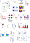
Figure 1)
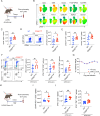
Figure 1). Generation of a CD28A210P ‘humanized’ mouse that better recapitulates the human response to CD28 superagonist antibody
A) Phylogenetic tree of CD28 C-terminal proline-rich domain amino acid sequence in tetrapod mammals, generated from uniprot sequence database(62). B) CD28A210P mice were generated by CRISPR-mediated substitution of proline for alanine at amino acid position 210 of mouse CD28. Gene and amino acid sequence of WT and CD28A210P CD28. C-E: Thymus of adult CD28A210P and WT mice assessed by flow cytometry for T cell development stage subsets (C), thymic FoxP3+ Tregs (D) and surface CD5 expression (E). F-H: Peripheral LN and spleen of adult WT and CD28A210P mice were assessed by flow cytometry for proportions of T cell subsets (F), Foxp3+ Tregs (G) and surface expression of CD28 (H). I-M: Mice were injected with CD28 superagonist (CD28SA) and monitored for weight loss (J), on day 4 post-injection cytokines were assessed in serum (K), and spleen analyzed by flow cytometry for frequencies and absolute counts of Foxp3+ Tregs (L) and activated CD4+ and CD8+ T cells (M). Data are pooled from at least two experiments with 3–6 per group, except E&H are one experiment representative of two, dots show individual mice, significance assessed by Student’s t-test or one-way ANOVA, * = p < 0.05, ** = p < 0.01, *** = p < 0.001.
Figure 2)
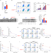
Figure 2). Enhancing CD28 C-terminal signaling increases initial effector T cell differentiation during acute and chronic viral infection
A) Experimental timeline for LCMV Armstrong infection and splenocyte analysis 7 days post infection. B) UMAP clustering and marker heatmap overlay of CD4+ and CD8+ cells by flow cytometry. Data pooled from WT and CD28A210P splenocytes to construct representative clustering. C) Absolute numbers of CD44+ CD8+ splenocytes. D) Representative gating and quantification of H-2Db GP33+ CD8+ T cells. E) Absolute numbers of effector CX3CR1+ and KLRG1+ CD8+ T cells. F) Representative gating and absolute numbers of cytokine producing CD8+ T cells following 4-hour ex vivo stimulation with LCMV peptide GP33. G) Weight loss following LCMV Armstrong infection shown as percentage of starting weight on day 0. H) Experimental timeline of LCMV clone 13 infection experiments. I) Absolute numbers of activated (CD44+ PD-1+) CD8+ T cells 8 days post infection with LCMV clone 13. J) Absolute numbers of IFNγ and IFNγ TNF producing CD8+ T cells following 4-hour ex vivo stimulation with LCMV peptide GP33. Data are pooled from at least two experiments with 4–8 per group. Significance assessed by Student’s t-test or one-way ANOVA, * = p < 0.05, ** = p < 0.01, *** = p < 0.001.
Fig 3)
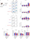
Fig 3). Enhancing CD28 C-terminal signaling induces early upregulation of CD8+ T cell inhibitory receptors and expedites exhaustion
A) Experimental timeline of LCMV clone 13 infection. B) Absolute numbers of splenic PD-1+ Tim-3+ CD8+ T cells 8 days post LCMV clone 13 infection. C) PD-1 GMFI of PD-1+ Tim-3+ CD8+ cells 8 days post infection. D) Serum IFNγ 8 days post infection. E) Ratio of CD8+ Teff (CX3CR1+) to Tex (CX3CR1 PD-1+ Tim-3+) splenocytes. F) Weight loss following LCMV infection, shown as a percentage of starting weight on day 0, data pooled from multiple experiments, (WT n= 9–28/time point) (CD28A210P n= 8–25/time point) student’s t test used for each day. G) Representative gating and quantification of absolute numbers of CD8+ effectors (CX3CR1+) and exhausted cells (CX3CR1 PD-1+ Tim-3+) on day 18 post infection. H) Ratio of absolute numbers of CD8+ CX3CR1+ effectors/ CD8+ CX3CR1 PD-1+ Tim-3+ exhausted cells. I) Representative gating and quantification of absolute numbers of IFNγ+ CD8+ T cells of indicated subsets based on previously described surface marker gating. Splenocytes were stimulated ex vivo for 4 hours with LCMV GP33 peptide in the presence of GolgiPlug followed by FACS staining. J) RT-qPCR quantification of ifng from LCMV clone 13 infected mouse kidney 18 days post infection. K) RT-qPCR quantification of viral copies in Kidney tissue from LCMV clone 13 infected mice 18 days post infection. Data are pooled from at least two experiments with 3–6 per group, except C and D one experiment representative of two and J is one experiment. Dots show individual mice. Significance assessed by Student’s t-test or one-way ANOVA, * = p < 0.05, ** = p < 0.01, *** = p < 0.001.
Figure 4)
Figure 4). The CD28 A210P mutation enhances MEK-dependent induction of JunB, IL-2, and PD-1/Tim-3 following T cell activation.
A-J) CD8+ T cells cells were stimulated with plate-bound anti-CD3 and anti-CD28 agonistic antibodies (1μg/mL in A,B E-J) (5μg/mL in C,D). A) Supernatant IL-2 was quantified by ELISA following 6 hours of stimulation. Each point represents the mean of replicate wells from an independent experiment. Data normalized to WT control in each independent experiment. B) Representative gating and quantification of PD-1 and Tim-3 on CD8+ cells stimulated for 2–3 days. Day 2: points indicate replicate wells pooled from 3 independent experiments. Day 3: points indicate replicate wells pooled from 2 independent experiments. C) Representative immunoblot of JunB from WT or CD28A210P CD8+ T cell nuclear extracts following stimulation for indicated times. Representative of 5 independent experiments. D) Representative immunoblot of JunB from nuclear extracts from WT or CD28A210P CD8+ T cells stimulated 2 hours in the presence of DMSO vehicle control or indicated concentrations of the MEK1/2 inhibitor, trametinib. Representative of 3 independent experiments. Quantification of vehicle treated extracts is pooled from 9 independent experiments and inhibitor treated extracts are pooled from 4 independent experiments. E) CD8+ cells were stimulated for 6 hours in the presence of vehicle or indicated concentrations of the MEK1/2 inhibitor. Supernatant IL-2 was quantified by ELISA. Data normalized to WT control in each independent experiment. Points indicate mean of pooled experimental replicates from each experiment. F) CD8+ cells were stimulated for 24 hours. Vehicle or MEK1/2 inhibitor were added to wells prior to cell seeding. Supernatant IFNγ was quantified by ELISA. Data normalized to WT control in each independent experiment. Points indicate mean of pooled experimental replicates from each experiment. G) Representative gating and quantification of PD-1 and Tim-3 on CD8+ cells stimulated for 24 hours with vehicle or MEK1/2 inhibitor added to wells prior to cell seeding. Data normalized to WT control in each independent experiment. Points indicate mean of pooled replicates from 2 independent experiments. H) CD8+ T cells were stimulated for 6 hours. Vehicle or indicated concentrations of the JNK1/2 inhibitor, JNK-IN-8, were added to wells prior to cell seeding. Supernatant IL-2 was quantified by ELISA. Data normalized to WT control in each independent experiment. Points indicate mean of pooled experimental replicates from 3 independent experiments. I) CD8+ cells were stimulated for 24 hours. Vehicle or JNK1/2 inhibitor were added to wells prior to cell seeding. Supernatant IFNγ quantified by ELISA. Data normalized to WT control in each independent experiment. Points indicate mean of pooled experimental replicates from 3 independent experiments. J) Representative gating and quantification of PD-1 and Tim-3 on CD8+ T cells stimulated 24 hours with vehicle or JNK1/2 inhibitor added prior to cell seeding. Data normalized to WT control in each independent experiment. Points indicate mean of pooled replicates from 2 independent experiments. Data are pooled or representative of at least two experiments with significance assessed by Student’s t-test or one-way ANOVA, * = p < 0.05, ** = p < 0.01, *** = p < 0.001.
Figure 5)
Figure 5). Enhancing CD28 signaling does not affect numbers of effector/exhausted T cells during late chronic infection
A) Experimental timeline of LCMV clone 13 infection with CD4-depletion. Anti-CD4 (GK1.5) was given I.P. (250 μg/mouse) on days −1 and +1. For experiments with anti-PD-L1 (10F.9G2) administration, 5 injections of anti-PD-L1 were administered I.P. (200 μg/injection) every 3 days for 2 weeks prior to analysis. B) Representative gating and quantification of frequencies and absolute counts of splenic CD8+ cell subsets on day 35+ of LCMV clone 13 infection. CD8+ subsets: effector (CX3CR1+) exhausted (CX3CR1 PD-1+ Tim-3). C) Representative gating and quantification of frequencies and absolute counts of IFNγ+ splenic CD8+ cell subsets on day 35+ of LCMV clone 13 infection. CD8+ subsets: effector (CX3CR1+) exhausted (CX3CR1 PD-1+ Tim-3). Splenocytes were re-stimulated ex vivo with LCMV GP33 peptide for 4 hours in the presence of GolgiPlug, followed by FACS staining. Data pooled from 2 independent experiments. D) Ratio of CD8+ effector (CX3CR1+) to exhausted (CX3CR1 PD-1+ Tim-3+) splenocytes 35+ days post LCMV clone 13 infection. E) RT-qPCR quantification of viral copies in Kidney, liver, and lung tissue from LCMV clone 13 infected mice 35+ days post infection. Data are pooled from two independent experiments with 3–5 per group, dots show individual mice, significance assessed by Student’s t-test or one-way ANOVA, * = p < 0.05, ** = p < 0.01, *** = p < 0.001.
Fig 6)
Fig 6). Enhancing CD28 increases Tpex differentiation and long-term maintenance without compromising memory precursors
A) Experimental timeline of LCMV Armstrong or clone 13 infection with CD4-depletion. Anti-CD4 (GK1.5) was given to indicated mice I.P. (250 μg/mouse) on days −1 and +1. B) Representative gating and quantification of absolute numbers of CD8+ Tpex (PD-1+ SLAMF6+ Tim-3) 18 days post infection. For assessment of IFNγ potential, splenocytes were stimulated with LCMV GP33 peptide ex vivo for 4 hours in the presence of GolgiPlug. C) Experimental timeline of LCMV clone 13 infection with CD4-depletion. Anti-CD4 (GK1.5) was given I.P. (250 μg/mouse) on days −1 and +1. For mice treated with anti-PD-L1 (10F.9G2), 5 injections of anti-PD-L1 were administered I.P. (200 μg/injection) every 3 days for 2 weeks prior to analysis. D) Representative histograms and GMFIs of CD28 on CD8+ Tpex (PD-1+ SLAMF6+ Tim-3−), effectors (CX3CR1+), and exhausted (CX3CR1 PD-1+ Tim-3+) cells 35+ days post LCMV infection. E) Representative gating and quantification of absolute counts of splenic CD8+ Tpex (PD-1+ SLAMF6+ Tim-3), effectors (CX3CR1+), and exhausted (CX3CR1 PD-1+ Tim-3+) cells 35+ days post LCMV infection. To determine absolute counts of IFNγ-producing CD8+ cells, 4-hour ex vivo LCMV GP33 peptide restimulation in the presence of GolgiPlug was performed. F) Quantification of absolute counts of splenic CD8+ Tpex (PD-1+ SLAMF6+ Tim-3) 8-, 18-, or 42- days post infection with LCMV clone 13 with or without anti-PD-L1 treatment for two weeks. G) Experimental timeline of LCMV Armstrong or clone 13 infection. Anti-CD4 (GK1.5) was given to indicated mice I.P. (250 μg/mouse) on days −1 and +1. H) Representative gating and absolute counts of splenic CD8+ Tpex (PD-1+ SLAMF6+ Tim-3−) 7–8 days post infection. I) Absolute counts of IFNγ-producing CD8+ Tpex (PD-1+ SLAMF6+ Tim-3−) following 4-hour ex vivo LCMV GP33 peptide restimulation in the presence of GolgiPlug. J) Representative histograms and GMFIs of PD-1 on Tpex following LCMV infection. K) Representative gating and quantification of absolute numbers of CD8+ SLECs (CD44+ KLRG1+ CD127−) and MPECs (CD44+ KLRG1 CD127+). Data are pooled or representative of at least two experiments with 3–6 per group, dots show individual mice, significance assessed by Student’s t-test or one-way ANOVA, * = p < 0.05, ** = p < 0.01, *** = p < 0.001.

References

    1. Burr J. S. et al., Cutting edge: distinct motifs within CD28 regulate T cell proliferation and induction of Bcl-XL. J Immunol 166, 5331–5335 (2001). - PubMed
    1. Dodson L. F. et al., Targeted knock-in mice expressing mutations of CD28 reveal an essential pathway for costimulation. Mol Cell Biol 29, 3710–3721 (2009). - PMC - PubMed
    1. Andres P. G. et al., Distinct regions in the CD28 cytoplasmic domain are required for T helper type 2 differentiation. Nat Immunol 5, 435–442 (2004). - PubMed
    1. Esensten J. H., Helou Y. A., Chopra G., Weiss A., Bluestone J. A., CD28 Costimulation: From Mechanism to Therapy. Immunity 44, 973–988 (2016). - PMC - PubMed
    1. Boomer J. S., Green J. M., An enigmatic tail of CD28 signaling. Cold Spring Harb Perspect Biol 2, a002436 (2010). - PMC - PubMed

Publication types