Case Reports
. 2025 Mar 28;2025(4):omaf011.
doi: 10.1093/omcr/omaf011. eCollection 2025 Apr.

Neuroendocrine carcinoma causing common bile duct obstruction: a case report

Affiliations
Case Reports

Neuroendocrine carcinoma causing common bile duct obstruction: a case report

Richard Shamoon et al. Oxf Med Case Reports. .

Abstract

A 46-year-old male with no comorbidities was referred to our hospital because of jaundice and elevated LFT markers. After further investigations, he underwent magnetic resonance cholangiopancreatography (MRCP), which revealed a hypo-enhancing periampullary mass measuring 15 mm in size causing common bile duct (CBD) dilatation of 12 mm in cross diameter with intrahepatic biliary obstruction, which explained the patient's symptoms. Side-view endoscopy was performed to obtain a specimen of the mass. Further histopathological workup revealed poorly differentiated neuroendocrine carcinoma (NEC). A multidisciplinary team (MDT) was conducted, and the patient was planned to undergo positron emission tomography-computed tomography (PET-CT) scan to investigate any further organ metastasis. Unfortunately, the patient missed his upcoming appointments and was lost to follow-up. Nevertheless, more research is needed to understand pathogenesis and the best course of management for small periampullary NETs.

Keywords: common bile duct obstruction; jaundice; periampullary carcinoma; periampullary neoplasms.


Conflict of interest statement

No conflicts of interest.

Figures
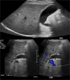
Figure 1
Figure 1
Abdominal axial ultrasound of the liver and gallbladder shows thick edematous gallbladder wall and a dilated common bile duct with intrahepatic biliary dilatation.
Figure 2
Figure 2
MRI diffusion/ADC map (A) shows restriction pattern of the periampullary lesion reflecting bright signal on diffusion and dark signal on ADC map. MRI axial T2 haste image at the level periampullary region (B) shows rounded isointense lesion. And post IV contrast axial MRI image (C) shows hypo enhancement of the peri-ampullary mass.
Figure 3
Figure 3
MRI coronal T2 haste (A) showing abrupt cut-off of the distal CBD with a dilated common bile duct, mild degree of intrahepatic biliary dilatation and mild dilatation of the pancreatic duct. MIP image (B) shows the cut off at the distal CBD, with severe dilatation of the extrahepatic and intrahepatic biliary tree, as well as dilated pancreatic duct.
Figure 4
Figure 4
Side view endoscopy showed an ulcerated ampullary mass.
Figure 5
Figure 5
Histopathological findings in H&E x 4 (A) shows variable sized nests of tumor in the lamina propria. H&E x 40 with mitosis (B) shows nuclear pleomorphism with frequent mitotic activity (black arrows). The tumor cells are strongly and diffusely positive for synaptophysin (C) and Ki-67 immunostain (D) displaying high proliferative index of more than 70%.

References

    1. Fernandes CJ, Leung G, Eads JR. et al. Gastroenteropancreatic neuroendocrine Tumors. Gastroenterol Clin N Am 2022;51:625–47. 10.1016/j.gtc.2022.06.002. - DOI - PubMed
    1. Hartel M, Wente MN, Sido B. et al. Carcinoid of the ampulla of Vater. J Gastroenterol Hepatol 2005;20:676–81. 10.1111/j.1440-1746.2005.03744.x. - DOI - PubMed
    1. Matli VVK, Wellman G, Jaganmohan S. et al. Ampullary and pancreatic neuroendocrine Tumors: a series of cases and review of the literature. Cureus 2022;14:e21657. 10.7759/cureus.21657. - DOI - PMC - PubMed
    1. Rindi G, Mete O, Uccella S. et al. Overview of the 2022 WHO classification of neuroendocrine neoplasms. Endocr Pathol 2022;33:115–54. 10.1007/s12022-022-09708-2. - DOI - PubMed
    1. Nagtegaal ID, Odze RD, Klimstra D. et al. The 2019 WHO classification of tumours of the digestive system. Histopathology 2020;76:182–8. 10.1111/his.13975 Epub 2019 Nov 13. - DOI - PMC - PubMed

Publication types

LinkOut - more resources