[Preprint]. 2025 Mar 12:rs.3.rs-6058394.
doi: 10.21203/rs.3.rs-6058394/v1.

At-Home Movement State Classification Using Totally Implantable Bidirectional Cortical-Basal Ganglia Neural Interface

Affiliations

At-Home Movement State Classification Using Totally Implantable Bidirectional Cortical-Basal Ganglia Neural Interface

Doris Wang et al. Res Sq. .

Abstract

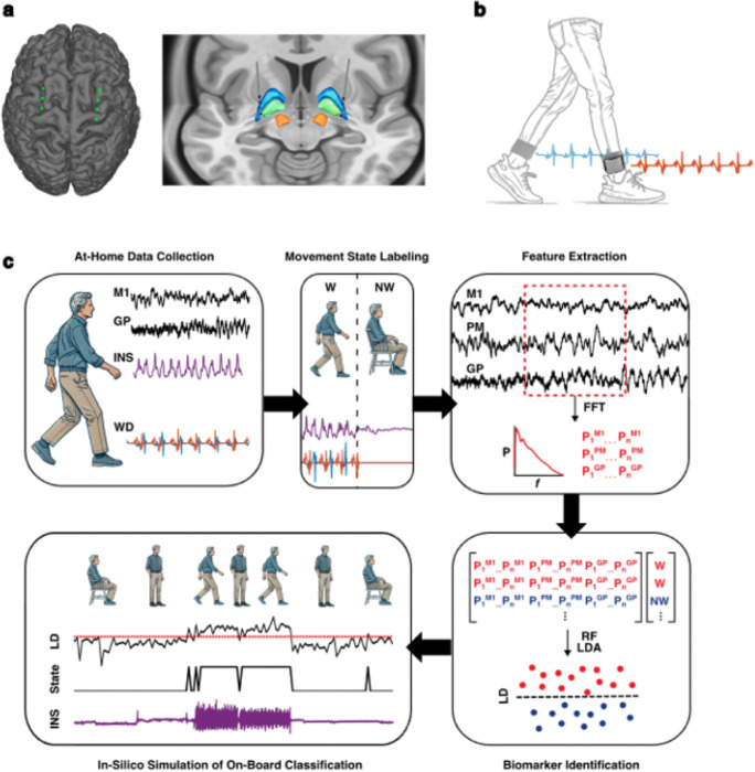
Movement decoding from invasive human recordings typically relies on a distributed system employing advanced machine learning algorithms programmed into an external computer for state classification. These brain-computer interfaces are limited to short-term studies in laboratory settings that may not reflect behavior and neural states in the real world. The development of implantable devices with sensing capabilities is revolutionizing the study and treatment of brain circuits. However, it is unknown whether these devices can decode natural movement state from recorded neural activity or accurately classify states in real-time using on-board algorithms. Here, using a totally implanted sensing-enabled neurostimulator to perform long-term, at-home recordings from the motor cortex and pallidum of four subjects with Parkinson's disease, we successfully identified highly sensitive and specific personalized signatures of gait state, as determined by wearable sensors. Additionally, we demonstrated the feasibility of using at-home data to generate biomarkers compatible with the classifier embedded on-board the neurostimulator. These findings offer a pipeline for ecologically valid movement biomarker identification that can advance therapy across a variety of diseases.

Keywords: Parkinson’s disease; deep brain stimulation; gait; globus pallidus; motor cortex; neurophysiology.


Conflict of interest statement

Competing Interests The authors declare no competing interests.

Figures

Figure 1:
Figure 1:. Pipeline for identification of cortical-pallidal neural biomarkers of at-home movement state in Parkinson’s disease.
a. Example localization of Summit RC+S bidirectional neurostimulator cortical electrodes overlying M1 and PM (left) and subcortical depth electrodes implanted in the GP (right). b. Representation of Rover accelerometer WDs, worn around the ankles bilaterally. Sample acceleration signals are shown from the left and right foot. c. Schematic representation of neural biomarker identification pipeline. Neural data from M1, PM, and GP and acceleration data from WD and INS were streamed from patients’ homes. Acceleration signals were aligned, partitioned into 10-second epochs, and labeled as periods of continuous walking (W) or non-walking (NW) using WD data. From each epoch, average power was calculated within all possible frequency bands from 1 to 50 Hz. These power value features were used to train and test LDA models. Finally, in-silico testing was performed to simulate continuous on-board classification of movement state using system-constrained biomarkers derived from at-home data. FFT = fast Fourier transform; GP = globus pallidus; INS = implantable neurostimulator; LD = linear discriminant; LDA = linear discriminant analysis; M1 = primary motor cortex; NW = non-walking; PM = premotor cortex; RF = random forest; W = walking; WD = wearable device.
Figure 2:
Figure 2:. Subject demographics and electrode localization.
a. Demographic and clinical characteristics are shown for the four subjects enrolled in this study. b. Subject-specific reconstructions are shown of ECoG contacts targeting M1 and PM and depth electrodes targeting GP. DBS = deep brain stimulation; ECoG = electrocorticography; GP = globus pallidus; M1 = primary motor cortex; MDS-UPDRS III = Movement Disorder Society – Unified Parkinson’s Disease Rating Scale (Third Revision); PIGD = postural instability and gait disorder; PM = premotor cortex.
Figure 3:
Figure 3:. Validation of wearable device (WD) accuracy in labeling walking and non-walking epochs.
a. Acceleration signals from L INS and bilateral WD are shown aligned with force signals from FSRs on the patient’s feet during a 300 second session of overground walking with interspersed periods of standing or seated rest. The bar at the top indicates patient behavior throughout the session (blue: walking; red: non-walking). A 5 second period of continuous walking indicated by the gray box is enlarged and displayed within the subpanel on the right. b. Subject-specific accuracies of WD-based movement state labeling are shown (Supplementary Figure S1b). FSR = force-sensitive resistor; HST = heel strike threshold; INS = implantable neurostimulator; LH = left heel; LT = left toe; RH = right heel; RT = right toe; WD = wearable device.
Figure 4:
Figure 4:. Spectral analysis of at-home walking and non-walking epochs.
a. Sample at-home recording is shown from Subject 4, with aligned acceleration signals from left WD (top) and INS (bottom). Periods of walking (blue) and non-walking (red) identified with WD-based labeling are indicated with shading. b. Sample mean PSDs are shown from 0 to 50 Hz for all walking (blue) and non-walking (red) 10-second epochs analyzed from GP, M1, and PM for Subject 4. c. Violin plots are shown comparing mean power within each canonical frequency band between all 10-second non-walking (red) and walking (blue) epochs. Two-sided Wilcoxon rank sum tests were used, with Benjamini-Hochberg correction for multiple comparisons. Across all hemispheres, M1 ⊠ and β power was significantly lower during walking epochs, highlighted with yellow boxes. * p < 0.05 (Supplementary Table S4). d. Normalized mean feature coefficients from logistic regression classifiers of movement state are visualized for all hemispheres. δ = delta (1–4 Hz); θ = theta (4–8 Hz); ⊠ = alpha (8–13 Hz); β = beta (13–30 Hz); γlow = low gamma (30–50 Hz); GP = globus pallidus; INS = implantable neurostimulator; M1 = primary motor cortex; PM = premotor cortex; WD = wearable device.
Figure 5:
Figure 5:. Identification of subject-specific movement biomarkers.
a. Normalized feature importance is shown for all cortical-pallidal frequency bands using mean MDI from 1,000 RF iterations. b. LDA movement state classifier performance is shown for each hemisphere and type of model (single-region, multi-region, and complete). Black dots indicate permuted chance-level performance (n = 1,000), which were used to calculate one-sided empirical p-values. * p < 0.001 (Supplementary Table S6). GP = globus pallidus; LDA = linear discriminant analysis; M1 = primary motor cortex; MDI = mean decrease in impurity; PM = premotor cortex; RF = random forest.
Figure 6:
Figure 6:. In-silico simulation of on-board movement state classification.
a. Performance of simulated on-board LDA classifiers is shown for models tested on at-home data (blue) and validated with testing on in-lab observed trials of overground walking with interspersed periods of standing or seated rest (orange). Empirical one-sided p-values were calculated by comparing model performance with a permuted chance-level distribution (n = 1,000) (Supplementary Table S7). Models deemed statistically insignificant (p ≥ 0.05) are shown with grayed bars. b. Sample in-silico simulation of continuous on-board classification using 5-second epochs is shown for Subject 1 using biomarker derived from at-home data. An observed trial is shown with true movement state indicated by the bar at the top. Simulated LD output is shown with the threshold indicated by red line, followed by the corresponding aDBS state and true INS acceleration signal. aDBS = adaptive deep brain stimulation; LD = linear discriminant; NW = non-walking; W = walking.

Similar articles

References

    1. Metzger SL, Littlejohn KT, Silva AB et al. (2023) A high-performance neuroprosthesis for speech decoding and avatar control. Nature 620(7976):1037–1046. 10.1038/s41586-023-06443-4 - DOI - PMC - PubMed
    1. Cervera MA, Soekadar SR, Ushiba J et al. (2018) Brain-computer interfaces for post-stroke motor rehabilitation: a meta-analysis. Ann Clin Transl Neurol 5(5):651–663. 10.1002/acn3.544 - DOI - PMC - PubMed
    1. Lorach H, Galvez A, Spagnolo V et al. (2023) Walking naturally after spinal cord injury using a brain–spine interface. Nature 618(7963):126–133. 10.1038/s41586-023-06094-5 - DOI - PMC - PubMed
    1. Willsey MS, Shah NP, Avansino DT et al. (2025) A high-performance brain–computer interface for finger decoding and quadcopter game control in an individual with paralysis. Nat Med 31(1):96–104. 10.1038/s41591-024-03341-8 - DOI - PMC - PubMed
    1. Neumann W, Gilron R, Little S, Tinkhauser G (2023) Adaptive Deep Brain Stimulation: From Experimental Evidence Toward Practical Implementation. Mov Disord 38(6):937–948. 10.1002/mds.29415 - DOI - PubMed

Publication types

LinkOut - more resources