[Preprint]. 2025 Mar 13:rs.3.rs-6097445.
doi: 10.21203/rs.3.rs-6097445/v1.

ACE2 utilization of HKU25 clade MERS-related coronaviruses with broad geographic distribution

Affiliations

ACE2 utilization of HKU25 clade MERS-related coronaviruses with broad geographic distribution

Chen Liu et al. Res Sq. .

Abstract

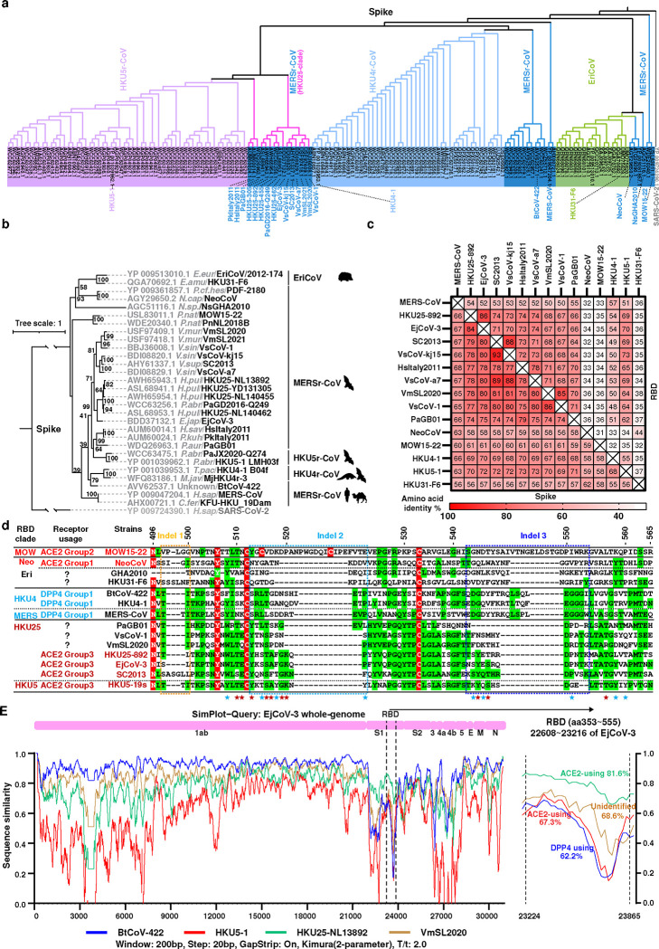
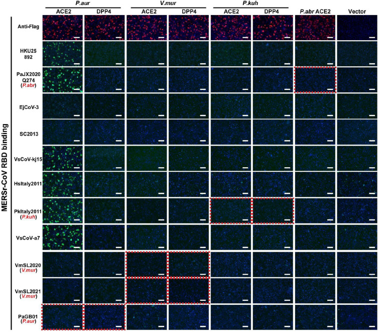
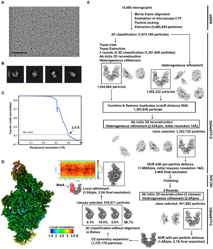
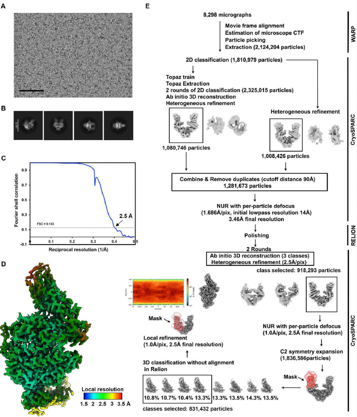
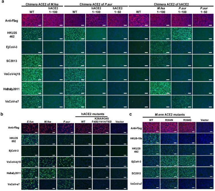
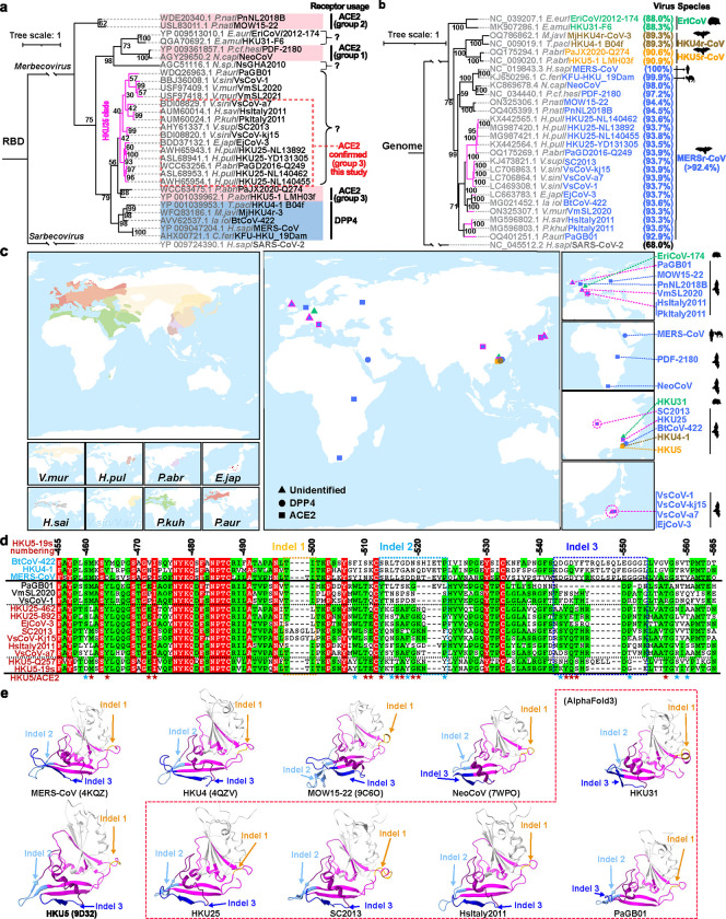
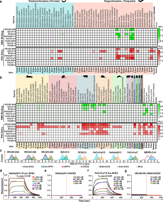
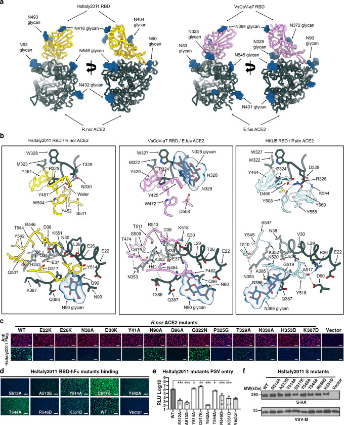
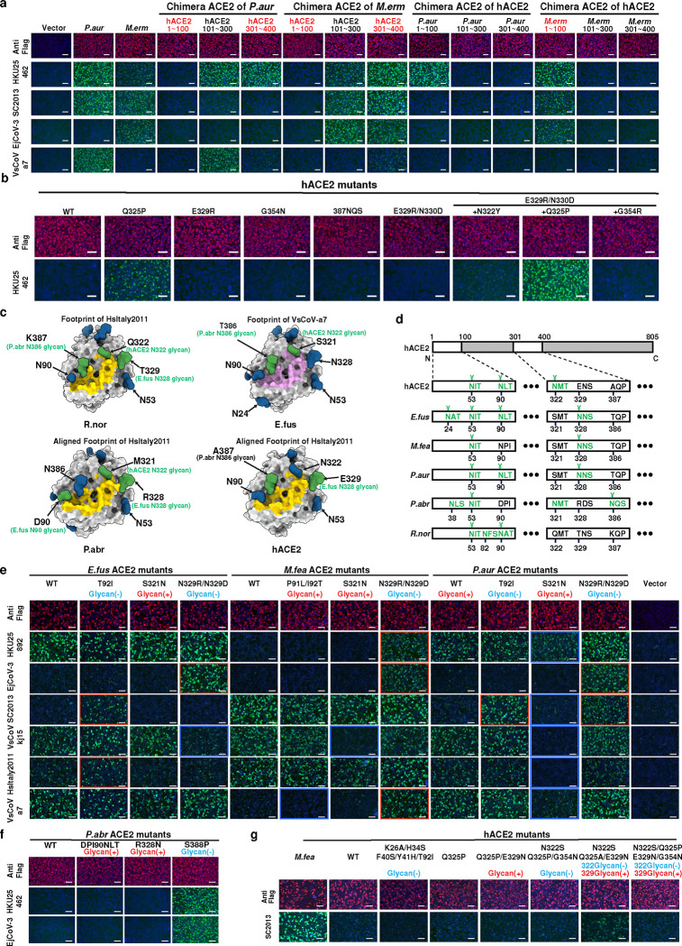
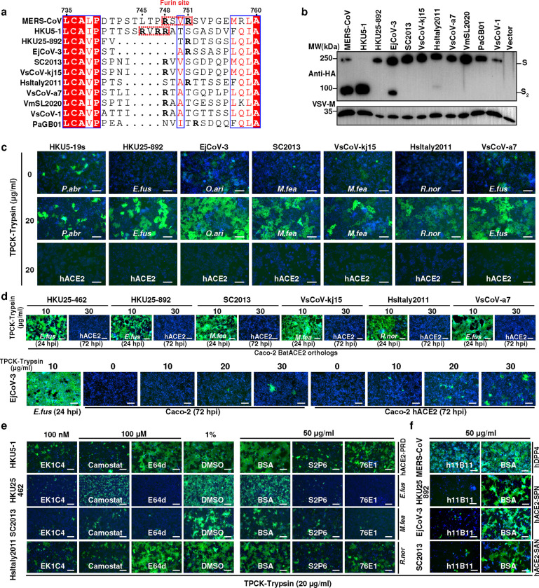
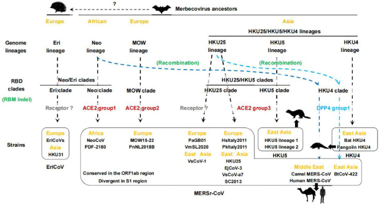
Dipeptidyl peptidase-4 (DPP4) is a well-established receptor for several MERS-related coronaviruses (MERSr-CoVs) isolated from humans, camels, pangolins, and bats 1-6. However, the receptor usage of many genetically diverse bat MERSr-CoVs with broad geographical distributions remains poorly understood. Recent studies have identified angiotensin-converting enzyme 2 (ACE2) as an entry receptor for multiple merbecovirus clades. Here, using viral antigen and pseudovirus-based functional assays, we demonstrate that several bat merbecoviruses from the HKU25 clade previously thought to utilize DPP4 7, employ ACE2 as their functional receptor. Cryo-electron microscopy analysis revealed that HsItaly2011 and VsCoV-a7 recognize ACE2 with a binding mode sharing similarity with that of HKU5 but involving remodeled interfaces and distinct ortholog selectivity, suggesting a common evolutionary origin of ACE2 utilization for these two clades of viruses. EjCoV-3, a strain closely related to the DPP4-using MERSr-CoV BtCoV-422, exhibited relatively broad ACE2 ortholog tropism and could utilize human ACE2 albeit suboptimally. Despite differences in entry mechanisms and spike proteolytic activation compared to MERS-CoV, these viruses remain sensitive to several broadly neutralizing antibodies and entry inhibitors. These findings redefine our understanding of the evolution of receptor usage among MERSr-CoVs and highlight the versatility of ACE2 as a functional receptor for diverse coronaviruses.

Keywords: ACE2; Cryo-EM; DPP4; EjCoV-3; HKU25; MERSr-CoV; Receptor.


Conflict of interest statement

Declaration of interests The authors declare no competing interests.

Figures

Extended Data Fig.1
Extended Data Fig.1. Amino acid sequence analysis of merbecovirus S glycoproteins.
a,b, phylogenetic trees based on amino acid sequences of S glycoproteins from all retrieved non-redundant merbecoviruses (a) or selected representative merbecoviruses S (b) were generated by IQ-tree2. SARS-CoV-2 was set as an outgroup. c, Heat plot of pairwise RBD and S amino acid sequence identities of indicated merbecoviruses. d, Manually adjusted multiple sequence alignment of RBM residues 496–565 (HKU5–19s residue numbering) from the indicated merbecoviruses with the three indels marked by dashed boxes. Fully and partially conserved residues were highlighted with red and green background, respectively. e, SimPlot analysis of whole-genome nucleotide similarity of indicated merbecoviruses relative to EjCoV-3. The right panel magnifies the RBD region (EjCoV-3 positions 22608~23216 nt). Dashed lines indicate RBD boundaries; nucleotide identities to EjCoV-3 are labeled.
Extended Data Fig.2
Extended Data Fig.2. Binding of HKU25 clade RBD-hFc constructs to ACE2 or DPP4 orthologs from selected bat host species.
HKU25-clade coronaviruses RBD-hFc binding to HEK293T cells transiently expressing the indicated receptors assessed by immunofluorescence. The Red dashed boxes highlight data that shows the RBD binding of indicated viruses with receptors from their reported host species (marked in red). Scale bars: 100 μm.
Extended Data Fig.3
Extended Data Fig.3. The expression level of ACE2 orthologs from various mammalian species.
a,b,Immunofluorescence analysis of the expression of bat (a) or non-bat mammalian (b) ACE2 orthologs in HEK293T cells by detecting the C-terminal fused FLAG tags.Scale bars: 100 μm.
Extended Data Fig.4
Extended Data Fig.4. Cryo-EM data processing of the E.fus ACE2 bound VsCoV-a7 RBD data set.
a,b, Representative electron micrographs (a) and 2D class averages (b) of the E.fus ACE2-bound VsCoV-a7 RBD complex embedded in vitreous ice. Scale bars: 100 nm (a) and 130 Å (b). c, Gold-standard Fourier shell correlation curve of the E.fus ACE2-bound VsCoV-a7 RBD reconstruction. The 0.143 cutoff is indicated by a horizontal dashed line. d, Local resolution estimation calculated using cryoSPARC and plotted on the sharpened map. e, Data processing flowchart. CTF: contrast transfer function; NUR: non-uniform refinement. The angular distribution of particle images calculated using cryoSPARC is shown as a heat map.
Extended Data Fig.5
Extended Data Fig.5. Cryo-EM data processing of the R.nor ACE2 bound HsItaly2011 RBD data set.
a,b, Representative electron micrographs (a) and 2D class averages (b) of the R.nor ACE2-bound HsItaly2011 RBD complex embedded in vitreous ice. Scale bars: 100 nm (a) and 130 Å (b). c, Gold-standard Fourier shell correlation curve of the R.nor ACE2-bound HsItaly2011 RBD reconstruction. The 0.143 cutoff is indicated by a horizontal dashed line. d, Local resolution estimation of the R.nor ACE2-bound HsItaly2011 RBD reconstruction calculated using cryoSPARC and plotted on the sharpened map. e, Data processing flowchart. CTF: contrast transfer function; NUR: non-uniform refinement. The angular distribution of particle images calculated using cryoSPARC is shown as a heat map.
Extended Data Fig.6
Extended Data Fig.6. Molecular determinants of ACE2 host species tropism overlapping with HKU5.
a, Immunofluorescence assay analyzing HKU25r-CoV RBD binding to HEK293T cells transiently expressing ACE2 chimeras with indicated sequence swaps between hACE2 and P.aur ACE2 or M.fea ACE2. The expression levels were validated by detecting the C-terminal fused FLAG tags. b, HKU25 clade viruses RBD-hFc binding to HEK293T cells transiently expressing hACE2 mutants. c, HKU25 clade viruses RBD-hFc binding to HEK293T cells transiently expressing M.erm ACE2 mutants. Scale bars: 100 μm.
Fig. 1 |
Fig. 1 |. Genetic features and geographic distribution of HKU25 clade MERSr-CoVs.
a,b, Maximum-likelihood phylogenetic trees of representative merbecoviruses, generated using IQ-tree2. Trees are based on amino acid sequences of the RBD (a) or genomic nucleotide sequences (b), with SARS-CoV-2 as the outgroup. Information on receptor usage, binding mode, host, and viral species based on amino acid sequence identities of five replicase domains (3CLpro, NiRAN, RdRp, ZBD, and HEL1) for coronavirus taxonomy (MERSr-CoV defined as diverged by less than 7.6% identity to MERS-CoV, NC_019843.3) are annotated. The known ACE2-using viruses were classified according to the three distinct binding modes identified in the NeoCoV- (group 1), MOW15–22- (group 2), and HKU5-related (group 3) clades . The scale bars denote genetic distance (1 substitution per nucleotide/amino acid position). c, Geographic distributions of bat hosts (left) and sampling locations of merbecoviruses with annotated receptor usage (right). Data from the IUCN (International Union for Conservation of Nature) Red List were visualized using GeoScene Pro. Squares: ACE2-using; Circles: DPP4-using; Triangles: receptor-unidentified. Color coding is the same as panel 1B. HKU25 clade strains were outlined in magenta. Host abbreviations: V.mur/V.sup (Vespertilio murinus/V.superans), H.pul (Hypsugo pulveratus), E.jap (Eptesicus japonensis), H.sai (Hypsugo savii), V.sin (Vespertilio sinensis), P.kuh (Pipistrellus kuhlii), P.aur (Plecotus auritus), P.abr (Pipistrellus abramus). d, RBM sequence alignment of the indicated merbecoviruses with manual adjustment to optimize indel positioning. Fully and partially conserved residues were indicated as red and green backgrounds, respectively. Dashed boxes highlight indels. Residues involved in HKU5-ACE2 interactions are marked with stars; positions that are conserved or non-conserved compared to HKU25 clade viruses are colored in red and blue, respectively. HKU5–19s residue numbering is shown. e, Experimentally determined structures or AlphaFold3-predicted RBD structures of representative merbecoviruses. The putative RBMs are indicated in magenta and three featured indels described in panel D are labeled in orange (indel 1), light blue (indel 2), and dark blue (indel 3), respectively. Sequences between indel 2 and indel 3 are labeled in purple to facilitate observation.
Fig. 2 |
Fig. 2 |. ACE2 ortholog utilization of HKU25 clade MERSr-CoVs.
a,b, Heat map representing the magnitude of HKU25 clade RBD-hFc binding to (green) and VSV pseudovirus (PSV) entry into (red) HEK293T cells transiently expressing bat (A) or non-bat b, mammalian ACE2 orthologs. Mammalian orders are color-coded: Carnivora, Primates, Artiodactyla, Rodentia, Cetacea, Perissodactyla, Diprotodontia, Pholidota, Erinaceomorpha, Lagomorpha, Chiroptera. Data represent mean values (n = 3 biological replicates). PSVs were pretreatment with 100 μg ml−1 TPCK-treated trypsin (Try). c, Flow cytometry analysis of binding of HKU25 clade RBDs to HEK293T transiently expressing the indicated ACE2 or DPP4 orthologs. Grey: vector control. Dashed lines: background threshold. Data are means of three technical repeats from three tubes of cells. d,g, BLI analysis of binding kinetics of dimeric ACE2 ectodomains (R.nor ACE2 in panel d, E.fus ACE2 in panel f, and hACE2 in panel e/g) to immobilized RBD-hFc of indicated strains. Analysis was conducted using curve-fitting kinetic with global fitting (1:1 binding model).
Fig. 3 |
Fig. 3 |. Structural basis for HsItaly2011 and VsCoV-a7 RBD interaction with bat or rat (R.nor) ACE2 orthologs.
a, Ribbon diagrams in two orthogonal orientations of the cryo-EM structures of the R.nor ACE2 peptidase domain (green) bound to HsItaly2011 RBD (gold) and E.fus ACE2 peptidase domain (green) bound to VsCoV-a7 RBD (plum). b, Zoomed-in views and comparisons of the interface key interactions of the HsItaly2011 RBD/R.nor ACE2, VsCoV-a7 RBD/E.fus ACE2 and HKU5 RBD/P.abr ACE2 (PDB ID: 9D32). HKU5 RBD and P.abr ACE2 peptidase domain are colored in light blue and green, respectively. Selected interface interactions are shown as black dotted lines. c, Analysis of HsItaly2011 RBD-hFc binding to membrane-anchored wildtype and mutants R.nor ACE2 transiently transfected in HEK293T cells analyzed by immunofluorescence. d, e RBD-hFc binding (d) and pseudovirus (pretreatment with 100 μg ml−1 TPCK-treated trypsin) entry (e) efficiencies of HsItaly2011 S mutants in HEK293T cells transiently expressing R.nor ACE2. f, VSV packaging efficiencies of HsItaly2011 S mutants. VSV-M was used as a loading control. Unpaired two-tailed t-tests for E, data are presented as means ± s.d. for for n = 3 biological replicates. *P < 0.05,**P < 0.01, ***P < 0.005. Scale bars in c and d: 100 μm. RLU: relative light unit.
Fig. 4 |
Fig. 4 |. ACE2 tropism determinants for HKU25 clade coronavirus.
a, Immunofluorescence analysis of RBD-hFc binding to HEK293T cells transiently expressing ACE2 chimeras (swaps between hACE2/P.aur ACE2 or hACE2/M.erm ACE2. ACE2 expression were validated by detecting the C-terminal fused FLAG tags. b, HKU25-NL140462 RBD-hFc binding to hACE2 mutants with equivalent residues in P.abr ACE2. c, N-glycans proximal to or within the HKU25 clade RBD-ACE2 interface (residues 1–100, 301–400). HsItaly2011 (yellow) and VsCoV-a7 (pink) RBD footprints are mapped onto ACE2 orthologs (gray surface). Glycans actually present on the surface of indicated WT ACE2 orthologs or predicted glycans through glycan-knock in mutations (based on hACE2, PDB 6M0J) are rendered in blue and green, respectively. d, Glycosylation sequons (green) at positions 53, 90, 322, 329, and 387 (hACE2 numbering). Cryo-EM confirmed glycans are marked with ¥. Please note although several glycosylation sequons are present, no glycan is present in these sites according to the cryo-EM map. e,g, RBD-hFc binding assay evaluating the impact of N-Glycan mutations on E.fus/M.fea/P.aur ACE2 (e), P.abr ACE2 (f) or hACE2 (g) orthologs. Red/blue dashed outlines: enhanced/reduced binding. Scale bars: 100 μm.
Fig. 5 |
Fig. 5 |. The Characterization and inhibition of the ACE2-mediated entry of rcVSV pseudotypes with HKU25 clade S glycoproteins.
a, S1/S2 junction sequence alignment of HKU25 clade S glycoproteins with MERS-CoV residue numbering. The arginines (R) were highlighted in bold fronts. Furin cleavage sites are highlighted in red dashed boxes. b, S glycoprotein incorporation into VSV pseudoviral particles by detecting the C-terminal fused HA tags. VSV-M was used as a loading control. c, Cell-cell fusion mediated by HKU25 clade coronaviruses S in Caco-2 cells stably expressing indicated ACE2 orthologs with the treatment of TPCK-treated trypsin. d, Propagation of rcVSV-HKU25r-S in Caco-2 cells or Caco-2 hACE2 cells in the presence of the indicated concentrations of TPCK-treated trypsin. e,f,Inhibition of rcVSV-HKU25r-S propagation by small molecular inhibitors, S2 antibodies (e) or hACE2-targeting antibodies h11B11 (f) in Caco-2 cells stably expressing indicated hACE2 mutants. BSA: Bovine serum albumin, 50 μg ml−1. hpi: hours post-infection. Scale bars: 200 μm.
Fig. 6 |
Fig. 6 |. Proposed evolutionary model of merbecovirus receptor usage acquisition and switching.
Geographical regions (orange) and receptor binding modes of specific merbecovirus RBD clades (gray: unidentified; red: ACE2; blue: DPP4) are indicated. Genome lineage evolution and RBD clade diversification involve RBM indels and S1 recombination (green) are annotated. Light and blue dashed lines propose the origins of DPP4-using MERS-CoV and BtCoV-422. Strains of the same species are grouped within black boxes. Spillovers to non-bat mammalian species are indicated. Indel: sequence insertions and deletions.

Similar articles

References

    1. Tse L. V. et al. A MERS-CoV antibody neutralizes a pre-emerging group 2c bat coronavirus. Sci Transl Med 15, eadg5567 (2023). - PMC - PubMed
    1. Wang Q. et al. Bat origins of MERS-CoV supported by bat coronavirus HKU4 usage of human receptor CD26. Cell Host Microbe 16, 328–337 (2014). - PMC - PubMed
    1. Chen J. et al. A bat MERS-like coronavirus circulates in pangolins and utilizes human DPP4 and host proteases for cell entry. Cell 186, 850–863.e16 (2023). - PMC - PubMed
    1. Raj V. S. et al. Dipeptidyl peptidase 4 is a functional receptor for the emerging human coronavirus-EMC. Nature 495, 251–254 (2013). - PMC - PubMed
    1. Lu G. et al. Molecular basis of binding between novel human coronavirus MERS-CoV and its receptor CD26. Nature 500, 227–231 (2013). - PMC - PubMed

Publication types

LinkOut - more resources