

This is a preprint.
Morphological and chemical characterization of nanoplastics in human tissue
- PMID: 40162223
- PMCID: PMC11952660
- DOI: 10.21203/rs.3.rs-6166886/v1
Morphological and chemical characterization of nanoplastics in human tissue
Abstract
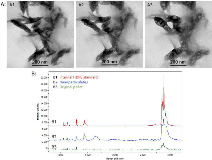
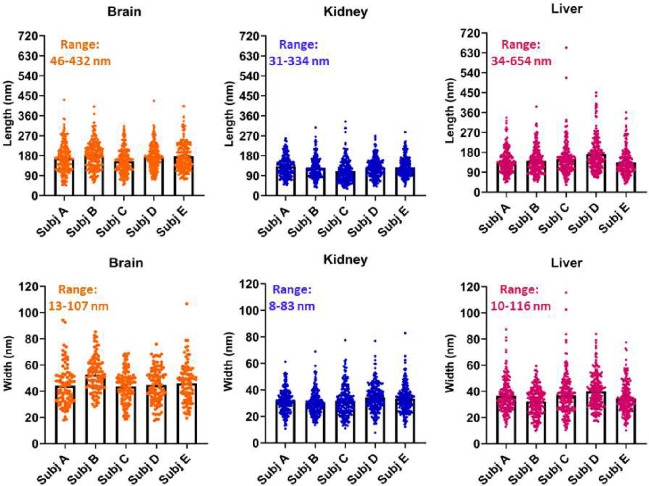
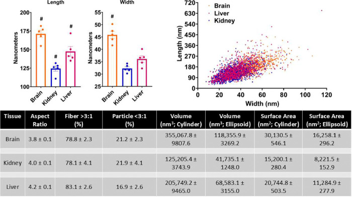
Micro- (≤ 5 mm) and nano- (≤ 1 μm) plastics have become ubiquitous resulting in inevitable human exposure. Evidence exists of mass-based accumulation of plastic in human tissues with visualization of micron-sized particles (> 1 μm). To date, there is little evidence to address accumulated nanoplastics. Understanding internalized plastic particle morphological and chemical characteristics is essential to facilitate proper design of future mechanistic and controlled exposure health effects studies to determine whether any health-related risks exist. Here we show microscopic evidence and quantitative dimensional analysis of nanoplastics in human decedent brain, kidney, and liver tissues. Mean particle lengths (nm) across the five decedents were 171.2±4.6 for brain, 124.4±3.6 for kidney, and 147.6±6.6 for liver. Mean particle widths (nm) were 45.9±1.5 for brain, 32.3±0.7 for kidney, and 36.1±1.3 for liver. When examining the aspect ratio, 78-83% consisted mostly of an elongated nanometer sized fiber morphology. The study provides isolation with physical and chemical characterization of nanoplastics in human tissues. Interestingly, differences were greater between tissues of a single decedent than across decedents. Consistently, the nanoplastics were largest in the brain. The observations overall suggest specificity with respect to systemic internalization and subsequent tissue accumulation of plastic particles less than one micron.
Keywords: bioaccumulation; brain; dimensions; kidney; liver; microplastics.
Conflict of interest statement
Competing Interests: The authors declare no competing financial and/or non-financial interests in relation to the work described.
Figures

Similar articles
-
Rapid single-particle chemical imaging of nanoplastics by SRS microscopy.Proc Natl Acad Sci U S A. 2024 Jan 16;121(3):e2300582121. doi: 10.1073/pnas.2300582121. Epub 2024 Jan 8. Proc Natl Acad Sci U S A. 2024. PMID: 38190543 Free PMC article.
-
Nano-scale and micron-scale plastics amplify the bioaccumulation of benzophenone-3 and ciprofloxacin, as well as their co-exposure effect on disturbing the antioxidant defense system in mussels, Perna viridis.Environ Pollut. 2024 Apr 1;346:123547. doi: 10.1016/j.envpol.2024.123547. Epub 2024 Feb 20. Environ Pollut. 2024. PMID: 38387549
-
Micro- and nanoplastics (MNPs) and their potential toxicological outcomes: State of science, knowledge gaps and research needs.NanoImpact. 2023 Oct;32:100481. doi: 10.1016/j.impact.2023.100481. Epub 2023 Sep 16. NanoImpact. 2023. PMID: 37717636 Free PMC article. Review.
-
Implications of exposure route for the bioaccumulation potential of nanopolystyrene particles.Chemosphere. 2024 Mar;351:141133. doi: 10.1016/j.chemosphere.2024.141133. Epub 2024 Jan 8. Chemosphere. 2024. PMID: 38199495
-
The Minderoo-Monaco Commission on Plastics and Human Health.Ann Glob Health. 2023 Mar 21;89(1):23. doi: 10.5334/aogh.4056. eCollection 2023. Ann Glob Health. 2023. PMID: 36969097 Free PMC article. Review.
References
-
- Ritchie H., Samborska V. & Roser M. Plastic pollution. Our world in data (2023).
-
- Organization W. H. Dietary and inhalation exposure to nano-and microplastic particles and potential implications for human health. (World Health Organization, 2022).
Publication types
Grants and funding
LinkOut - more resources
Full Text Sources
