[Preprint]. 2025 Mar 14:2025.03.13.25323904.
doi: 10.1101/2025.03.13.25323904.

Disease stage-specific atrophy markers in Alzheimer's disease

Hannah Baumeister  1 Helena M Gellersen  1   2   3 Sarah E Polk  1 René Lattmann  1   4 Anika Wuestefeld  5 Laura E M Wisse  6 Trevor Glenn  7 Renat Yakupov  1   4 Melina Stark  8   9 Luca Kleineidam  8   9 Sandra Roeske  8 Barbara Marcos Morgado  10 Hermann Esselmann  10 Frederic Brosseron  8 Alfredo Ramirez  8   9   11   12   13 Falk Lüsebrink  1 Matthis Synofzik  14   15   16 Björn H Schott  10   17   18 Matthias C Schmid  8   19 Stefan Hetzer  20 Peter Dechent  21 Klaus Scheffler  22 Michael Ewers  23   24 Julian Hellmann-Regen  25   26   27 Ersin Ersözlü  25   26   27 Eike Spruth  25   28 Maria Gemenetzi  25   28 Klaus Fliessbach  8   9 Claudia Bartels  10 Ayda Rostamzadeh  29 Wenzel Glanz  1 Enise I Incesoy  1   4   30 Daniel Janowitz  24 Boris-Stephan Rauchmann  31   32   33 Ingo Kilimann  34   35 Sebastian Sodenkamp  14   36 Marie Coenjaerts  8 Annika Spottke  8   37 Oliver Peters  25   28 Josef Priller  25   28   38   39   40 Anja Schneider  8   9 Jens Wiltfang  10   17   41 Katharina Buerger  23   24 Robert Perneczky  23   31   42   43 Stefan Teipel  34   35 Christoph Laske  14   36   44 Michael Wagner  8   9 Gabriel Ziegler  1   4 Frank Jessen  8   11   29 Emrah Düzel  1   4 David Berron  1   5   45 DELCODE study group
Affiliations

Disease stage-specific atrophy markers in Alzheimer's disease

Hannah Baumeister et al. medRxiv. .

Update in

  • Disease stage-specific atrophy markers in Alzheimer's disease.
    Baumeister H, Gellersen HM, Polk SE, Lattmann R, Wuestefeld A, Wisse LEM, Glenn T, Yakupov R, Stark M, Kleineidam L, Roeske S, Morgado BM, Esselmann H, Brosseron F, Ramirez A, Lüsebrink F, Synofzik M, Schott BH, Schmid MC, Hetzer S, Dechent P, Scheffler K, Ewers M, Hellmann-Regen J, Ersözlü E, Spruth E, Gemenetzi M, Fliessbach K, Bartels C, Rostamzadeh A, Glanz W, Incesoy EI, Janowitz D, Rauchmann BS, Kilimann I, Sodenkamp S, Coenjaerts M, Spottke A, Peters O, Priller J, Schneider A, Wiltfang J, Buerger K, Perneczky R, Teipel S, Laske C, Wagner M, Ziegler G, Jessen F, Düzel E, Berron D; DELCODE study group. Baumeister H, et al. Alzheimers Dement. 2025 Jul;21(7):e70482. doi: 10.1002/alz.70482. Alzheimers Dement. 2025. PMID: 40691867 Free PMC article.

Abstract

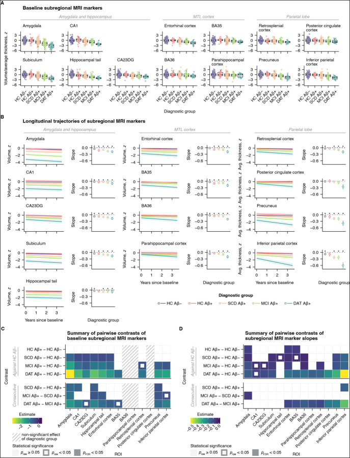
Introduction: Structural MRI often lacks diagnostic, prognostic, and monitoring value in Alzheimer's disease (AD), particularly in early disease stages. To improve its utility, we aimed to identify optimal MRI readouts for different use cases.

Methods: We included 363 older adults; healthy controls (HC) who were negative or positive for amyloidbeta (Aβ) and Aβ-positive patients with subjective cognitive decline (SCD), mild cognitive impairment, or dementia of the Alzheimer type. MRI and neuropsychological assessments were administered annually for up to three years.

Results: Accelerated atrophy of distinct MTL subregions was evident already during preclinical AD. Symptomatic disease stages most notably differed in their hippocampal and parietal atrophy signatures. Associations of atrophy markers and cognitive inventories varied by intended use and disease stage.

Discussion: With the appropriate readout, MRI can detect abnormal atrophy already during preclinical AD. To optimize performance, MRI readouts should be tailored to the targeted disease stage and intended use.


Conflict of interest statement

J.W. has been an honorary speaker for Beeijing Yibai Science and Technology Ltd, Eisai, Gloryren, Janssen, Pfizer, Med Update GmbH, Roche, Lilly, Roche Pharma, and has been a member of the advisory boards of Abbott, Biogen, Boehringer Ingelheim, Lilly, Immungenetics, MSD Sharp-Dohme, Noselab, Roboscreen, Roche Pharma and receives fees as a consultant for Immungenetics, Noselab, and Roboscreen. J.W. holds the following patents: PCT/EP 2011 001724 and PCT/EP 2015 052945. All other authors report no conflicts of interest relevant to this work.

Figures

Figure 1
Figure 1. Baseline and longitudinal MTL and parietal subregional structure across the clinical AD continuum.
(A) Boxplot diagrams of baseline gray matter volumes and average cortical thicknesses across regions of interest, grouped by meta-region. (B) Group-level longitudinal trajectories of structural markers (left panels) and the corresponding slopes (right panels), estimated from linear mixed-effects models. Ribbons and error bars denote 95% confidence intervals. Asterisks highlight significant slopes, i.e., significant change over time. (C) Matrix displaying estimates from ANCOVA post hoc group comparisons of baseline MRI markers. The striped pattern indicates measures with non-significant main effects of diagnostic group (not passed to post hoc tests). (D) Estimates from pairwise group comparisons of longitudinal slopes of subregional MRI markers. Abbreviations: BA, Brodmann area. CA, cornu ammonis. CA23DG, cornu ammonis 2, 3, and dentate gyrus. DAT, dementia of the Alzheimer type. FDR, false discovery rate. HC, healthy controls. IPC, inferior parietal cortex. MCI, mild cognitive impairment. MTL, medial temporal lobe. ROI, region of interest. SCD, subjective cognitive decline. SUB, subiculum.
Figure 2
Figure 2. Non-linear changes in MTL and parietal subregional structure along a continuous estimate of disease progression, scaled in years.
(A) Boxplot diagrams of baseline disease stage by diagnostic group, centered to the median of the HC Aβ+ group. (B) Cubic B-splines and their first derivatives were used to assess the development of structural MRI measures with disease progression. The estimated trajectories also shown in (C) with bootstrapped confidence intervals and clustered by meta-region. (D) For each ROI, the predicted curves also shown in (B) and (C) are plotted along with the observed participant-level trajectories. Abbreviations: Aβ, amyloid-β. AMY, amygdala. BA, Brodmann area. CA, cornu ammonis. CA23DG, cornu ammonis 2, 3, and dentate gyrus. DAT, dementia of the Alzheimer type. ERC, entorhinal cortex. HC, healthy controls. IPC, inferior parietal cortex. MCI, mild cognitive impairment. MTL, medial temporal lobe. PCC, posterior cingulate cortex. PHC, parahippocampal cortex. PRE, precuneus. ROI, region of interest. RSC, retrosplenial cortex. SCD, subjective cognitive decline. SUB, subiculum. TAIL, hippocampal tail.
Figure 3
Figure 3. Estimates from bivariate LGCMs capturing the disease stage-specific cross-sectional and longitudinal relationships of subregional structural MRI measures and cognitive test scores.
The covariance parameters of interest were the (A) baseline-baseline (B) baseline-slope, and (C) slope-slope pairwise associations of ROI structure and cognitive score. Abbreviations: AMY, amygdala. ADAS-Cog, Alzheimer’s Disease Assessment Scale-Cognitive subscale. BA, Brodmann area. CA, cornu ammonis. CA23DG, cornu ammonis 2, 3, and dentate gyrus. CSD-SB, Clinical Dementia Rating Sum of Boxes scale. ERC, entorhinal cortex. FAQ, Functional Activities Questionnaire. FCSRT, Free and Cued Selective Reminding Test. FDR, false discovery rate. IPC, inferior parietal cortex. MMSE, Mini-Mental State Examination. MTL, medial temporal lobe. PACC-5, Preclinical Alzheimer Cognitive Composite. PHC, parahippocampal cortex. PRE, precuneus. RSC, retrosplenial cortex. SUB, subiculum. TAIL, hippocampal tail.

References

    1. Jack CR, Andrews JS, Beach TG, et al. Revised criteria for diagnosis and staging of Alzheimer’s disease: Alzheimer’s Association Workgroup. Alzheimer’s Dement. 2024;20(8):5143–5169. doi: 10.1002/alz.13859 - DOI - PMC - PubMed
    1. Baumeister H, Vogel JW, Insel PS, et al. A generalizable data-driven model of atrophy heterogeneity and progression in memory clinic settings. Brain. 2024;147(7):2400–2413. doi: 10.1093/brain/awae118 - DOI - PMC - PubMed
    1. Bejanin A, Schonhaut DR, Joie RL, et al. Tau Pathology and Neurodegeneration Contribute to Cognitive Impairment in Alzheimer’s Disease. Brain. 2017;140(12):3286–3300. doi: 10.1093/brain/awx243 - DOI - PMC - PubMed
    1. Rafii MS, Aisen PS. Detection and treatment of Alzheimer’s disease in its preclinical stage. Nat Aging. 2023;3(5):520–531. doi: 10.1038/s43587-023-00410-4 - DOI - PMC - PubMed
    1. Hansson O. Biomarkers for Neurodegenerative Diseases. Nat Med. 2021;27(6):1–10. doi: 10.1038/s41591-021-01382-x - DOI - PubMed

Publication types

LinkOut - more resources