

Fenestration and Bifurcation of the Internal Jugular Vein; Surprises During Head and Neck Surgery
- PMID: 40162379
- PMCID: PMC11949429
- DOI: 10.22038/ijorl.2025.83514.3810
Fenestration and Bifurcation of the Internal Jugular Vein; Surprises During Head and Neck Surgery
Abstract
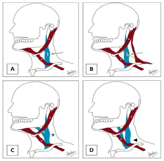
Introduction: The internal jugular vein (IJV) is one of the major vessels in the neck and serves as an important landmark for surgeons during head and neck surgery. Anomalies of the IJV are rare and seldom encountered by the surgeons. However, a comprehensive knowledge of these variations is essential for better surgical dissection and to prevent intra-operative mishaps. The variations can be in the forms of bifurcation, trifurcation, duplication, fenestration and posterior tributaries of the IJV. Here we describe three cases of bifurcation and fenestration of the IJV that we encountered in our surgical practice.
Case report: In the first patient, we found an empty fenestration of the right internal jugular vein during a selective neck dissection for tongue carcinoma. The spinal accessory nerve was passing lateral to the IJV above the level of the fenestration. The second patient was operated for a left vagal schwannoma in the neck. During the surgery, we found a bifurcation of the left IJV, and the two tributaries fused just above the left omohyoid muscle. The third patient, a sixty-year-old lady also had a bifurcation of the left IJV. It was found during a modified radical neck dissection for carcinoma ex pleomorphic adenoma of the left parotid gland.
Conclusion: An in-depth knowledge of the anomalies of the internal jugular vein and meticulous evaluation of the pre-operative imaging may help the surgeons in preventing any intra-operative catastrophe during head and neck surgery.
Keywords: Anatomical variation; Bifurcation; Fenestration; Internal jugular vein; Spinal accessory nerve.
©2025 Mashhad University of Medical Sciences.
Conflict of interest statement
The authors declare that there was no conflict of interest.
Figures


References
-
- Rivard AB, Kortz MW, Burns B. StatPearls (Internet) Treasure Island (FL): StatPearls Publishing; 2023 . Anatomy, Head and Neck: Internal Jugular Vein. - PubMed
-
- Ibrahim B, Berania I, Moubayed SP, Christopoulos A, Ayad T. Internal Jugular Vein Duplication: Anatomic Relationship With the Spinal Accessory Nerve. J Oral Maxillofac Surg. 2016 Jul;74(7):1502.e1. - PubMed
-
- Prades JM, Timoshenko A, Dumollard JM, Durand M, Merzougui N, Martin C. High duplication of the internal jugular vein: clinical incidence in the adult and surgical consequences, a report of three clinical cases. Surg Radiol Anat. 2002 May;24(2):129. - PubMed
Publication types
LinkOut - more resources
Full Text Sources